Researchers use multiplexed 3D imaging to perform molecular and morphological assessments of colorectal cancers
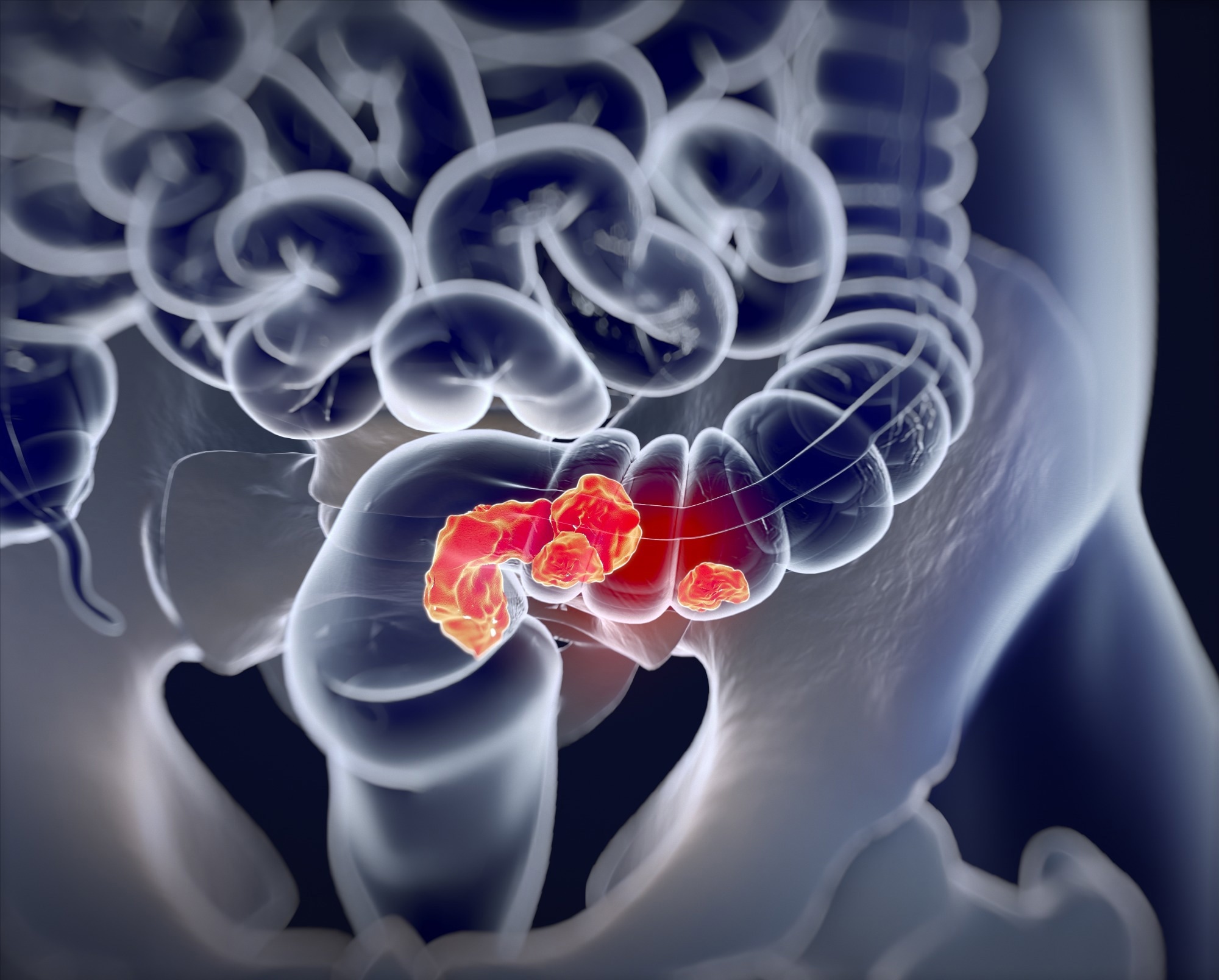
In a recent study published in Cell, researchers used multiplexed and three-dimensional imaging with machine-based learning and spatial statistics to perform molecular and morphological assessments of colorectal cancers (CRC).

Solid tumors, in the advanced stage, are complicated assimilations of tumor cells, stromal cells, and immunological cells with considerable histomorphological variations within the tumor. The conventional histopathological analysis provides inadequate data for precision medicine and mechanistic studies. Spatial tumor atlases can build on the histological foundation and contemporary tumor genetics by obtaining in-depth three-dimensional morphological and molecular data.
About the study
In the present study, researchers performed a novel highly-multiplexed three-dimensional tissue imaging analysis to construct CRC atlases providing data at subcellular resolution.
The images were segmented, and their fluorescence intensities were quantified for generating data at a singular-cell level on cellular type, state, and cell-cell interactions. Three-dimensional reconstructions of serial tissue sections were prepared and supervised by machine learning. Two approaches were used for analyzing tumor cells, i.e., a ‘bottoms-up’ approach and a ‘top-down’ approach.
The ‘bottoms-up’ approach involved using spatial statistics for enumerating cell type, assessing cellular interactions, and generating local neighbourhoods, leveraging tools used in single-cell analyses such as mass cytometry and single-cell ribonucleic acid sequencing (scRNA-seq). Contrastingly, the ‘top-down’ approach involved annotating histological features or histotypes related to disease states or outcomes, followed by multiplexed data computations for identifying the underlying patterns at a molecular level.
High-plex cyclic immunofluorescence analysis (CyCIF) and hematoxylin and eosin (H&E)-stained CRC images obtained by the histopathological analysis were combined with scRNA-seq analysis and microregion transcriptomic analysis. Further, the CyCIF images were analyzed by t-SNE (t-distributed stochastic neighbour embedding) and the transcriptomic microregions were analyzed by PCA (principal component analysis). In addition, a virtual tissue microarray (vTMA) analysis was performed.
Results
Multiplexed analysis showed intermixed tumor morphologies and molecular gradients. Various cancer-characteristic cellular features were observed as large, interconnected structures. Three-dimensional TLS (tertiary lymphoid structure) networks showed intra-TLS patterning variations. PD1 (programmed cell death protein 1)-PDL1 (programmed death ligand-1) interactions were observed primarily between T lymphocytes and myeloid cells in the colorectal cancer cohort.
Immunology eBook

The analyses showed recurrently occurring transitions between tumor morphologies, a few of which were coincident with extensive gradients in the epigenetic regulator and oncogene expression. At the tumor invasive margins, the sites of competition between cancerous cells, normal cells, and immunological cells, T lymphocyte counts were reduced, involving several types of cells. Apparently localized archetypical two-dimensional CRC characteristics like TLS were found to be interconnected with molecular gradients and considerably larger in the three-dimensional analysis.
The tumor microenvironment (TME) showed spatial organization spanning over three to four orders of magnitude. The tumor invasive margins showed budding-type invasive margins, mucinous invasive margins, and deeply extending pushing-type invasive margins, extending the tumor into the underlying connective tissue and smooth muscles. The cellular segmentations in 75 WSI images showed that CK+ (cytokeratin-positive) cells of the normal and tumor epithelium were separated from the cluster of differentiation 31+ (CD31+) cells of the endothelium, primarily blood vasculature, desmin-positive cells of stroma, and CD45-positive immunological cells.
Immunological cells observed within the tumor comprised CD8+ and programmed cell death protein 1-expressing cytotoxic-type T lymphocytes, CD4-expressing helper-type T lymphocytes, CD68-expressing and/or CD163-expressing macrophages, and CD20-expressing B lymphocytes. In addition, subcategories like CD4+ FOXP3+ Tregs (regulatory T lymphocytes) were observed. Solid adenocarcinomas showed the greatest percentage of cytokeratin-positive cancer cells (70.0%), whereas the adjacent normal epithelial tissues had the least cytokeratin-positive cells (25.0%) and showed immune cell and stromal cell abundance.
In colorectal cancer (CRC)1 to 17, correlation lengths observed ranged between 80.0 mm for the cluster of differentiation 31 positivity and 400.0 mm for CD20 or keratin positivity. The lengths were associated with recurrent histomorphological characteristics, inclusive of small-sized capillaries among the cluster of differentiation 31-positive cells, tumor sheets for cytokeratin-positive cell types, and tertiary lymphoid structures for the cluster of differentiation 20-positive cells.
The virtual TMA and real TMA scores were comparable, and the CyCIF findings matched theoretical data. In line with kNN classified modeling of CyCIF analysis data, graded transitions were observed from low-grade mucinous/glandular to high-grade budding/fragmented histologies among the tumor/epithelial and immunological/stromal tissue compartments. CyCIF markers showed graded intensities within an entire tumor or coinciding with localized morphology gradients.
The findings indicated that epithelial-mesenchymal-like transitions and TB in CRC1 were characterized by the formation of large fibrillar structures appearing as small-sized buds in cross-sections at the distal tips. Fibrils could invade different environments, including mucin and stroma, that seem to form by gradual disruption of cellular adhesion related to graded epithelial-mesenchymal-like transitions. The team observed less tumor proliferation at buds and greater proliferation at deep invasive tumor margins, with differing levels of phosphotyrosine pathway activation, and immunological suppression.
Overall, the study findings showed that multiplexed WSI analysis characterizes graded and mixed molecular and morphological characteristics in human CRC specimens, highlighting large-sized and distinctive structural characteristics and intra-TLS variations in spatial patterns of tumors.
- Jia-Ren Lin et al. (2023). Multiplexed 3D atlas of state transitions and immune interaction in colorectal cancer. Cell. doi: https://doi.org/10.1016/j.cell.2022.12.028 https://www.cell.com/cell/fulltext/S0092-8674(22)01571-9
Posted in: Medical Science News | Medical Research News
Tags: Adenocarcinomas, Blood, Cancer, Capillaries, CD4, Cell, Cell Death, Colorectal, Colorectal Cancer, Cytometry, Fluorescence, Genetics, Imaging, Ligand, Lymphocyte, Machine Learning, Medicine, Microarray, Morphology, Oncogene, Precision Medicine, Programmed Cell Death, Proliferation, Protein, Ribonucleic Acid, T Lymphocyte, Top-Down, Vasculature

Written by
Pooja Toshniwal Paharia
Dr. based clinical-radiological diagnosis and management of oral lesions and conditions and associated maxillofacial disorders.
Source: Read Full Article
