Study of natural killer lymphocytes transcriptionally reprogrammed by vaccinia virus
In a recent study posted to the bioRxiv* preprint server, researchers evaluated responses of natural killer (NK) lymphocytes to VACV (vaccinia virus) infections in vivo.
Studies have reported that NK lymphocytes play a crucial role in controlling poxvirus infections. Therefore, there is a growing scientific interest in exploiting their abilities in terms of poxvirus-based oncolytic therapies and vaccinations. However, data on the mechanisms of NK cell activation, receptors involved in recognizing VACV-infected lymphocytes, and the ways of reprogramming NK lymphocytes as a result of VACV infections are limited.
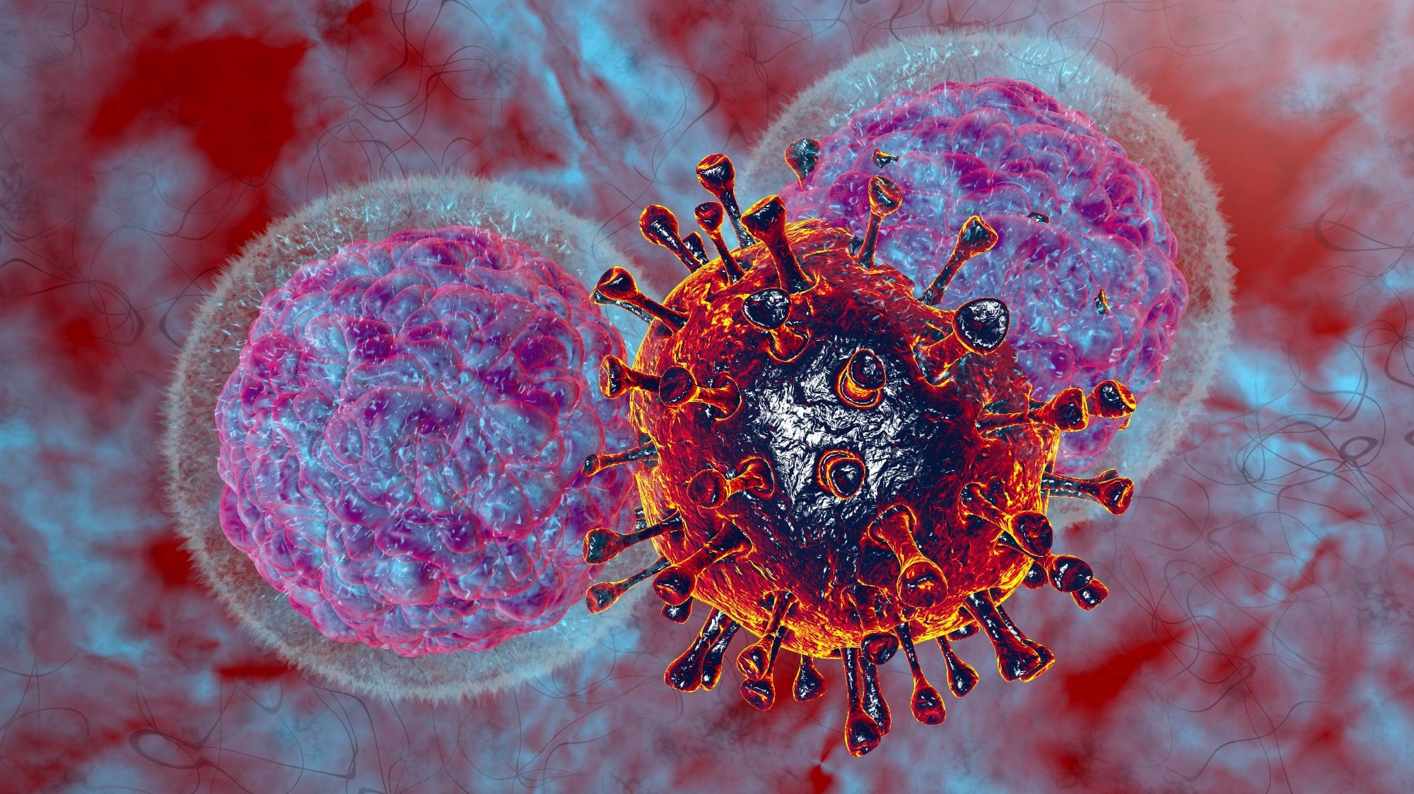
 Study: Transcriptional reprogramming of natural killer cells by vaccinia virus shows both distinct and conserved features with mCMV. Image Credit: Numstocker / Shutterstock
About the study
In the present study, the researchers at the University of Cambridge, UK, and the Fred Hutchinson Cancer Centre, USA, evaluated changes in NK lymphocyte transcriptomes in mice due to intranasal VACV WR (western reserve) strain infection.
Subsequently, the changes were compared to identical NK lymphocyte transcriptomic data from modified vaccinia Ankara (MVA)-infected humans and cytomegalovirus (mCMV)-infected C57BL/6 mice. Further, the transcriptional profiles of NK lymphocytes were compared with flow cytometry-determined cell surface molecule abundance. Fluorescence-activated cell sorting (FACS) analysis and lymphocyte isolation experiments were performed.
Ribonucleic acid (RNA) extracted from NK lymphocytes was subjected to bulk RNA sequencing (RNAseq) analysis. For assessing biological pathways affected by VACV infections in NK lymphocytes, differentially-expressed genes (DEG) between the samples of VACV-infected mice and mock animals were analyzed by biologic process gene ontologies enrichment analysis.
For the intranasally administered murine model, the animals were anesthetized and infected with 5.0 × 10³ PFU (plaque-forming units) of VACV WR strain for primary VACV infections or 105 PFU for the post-VACV vaccination challenge or were given control injections. For the intradermally administered murine infection model, the animals were intradermally infected with 103 PFU of the VACV WR strain. In addition, plaque assays were performed to confirm VACV titers in BSC-1 cells (African green monkey cell line).
Twenty-eight days post intradermal VACV infection, blood and spleen samples were obtained from mice. Splenic tissues were used for NK or cluster of differentiation 8+ (CD8+) T lymphocyte isolation. The tail veils of recipient murine animals were intravenously injected with NK lymphocytes, CD8+ T lymphocytes, or suspensions of splenocytes, and the mice were intranasally challenged with 105 PFU of VACV WR after one day. The animals were regularly weighed and observed for any signs of the disease up to 14 days post-VACV challenge.
Results
Broad alterations were observed in the activity of NK lymphocyte transcripts among VACV-infected mice in accordance with direct recognition of target cells and exposure to cytokines and interferons. In addition, the team noted alterations in the expression of particular NKRs (NK surface receptors), such as the SLAM and Ly49 family receptors, and memory-associated NK marker upregulation. Adoptive transfer of NK memory cells did not confer immune protection against reinfections. Comparing the findings with NK lymphocyte responses to murine cytomegalovirus (mCMV) infections showed common features and distinct VACV-induced-NK transcriptional activity. An overlap between NK transcriptional responses among humans administered attenuated VACV vaccines, and MVA was observed, indicative of conserved NK lymphocyte responses among different hosts. VACV-induced activation, expansion, and maturation of NK lymphocytes with simultaneous changes in the bulk transcriptional programs and memory marker expression.
NK cell numbers increased at 6.5 days post-infection (dpi), and preferential expansion of CD27+CD11b- and CD27+CD11b+ subsets was observed. DEG (differentially expressed genes) analysis between VACV-infected and mock samples, at 1.5 dpi and 6.5 dpi, respectively, showed 70 transcripts and 3280 transcripts with significant alterations. Comparing pairs of VAC-infected samples and mock samples showed 4045 and 140 significantly altered transcripts, respectively.
DEGs in VACV-infected murine animals at 1.5 dpi could not be distinguished from background noises, whereas those identified in the infected animals at 6.5 dpi differed biologically in their responses to VACV infections. NK lymphocytes engaged effector pathways and displayed active defense phenotypes resulting from VACV infections. NK lymphocytes not only recognized abnormal protein expression patterns at the surface of VACV-infected cells in vivo but also the interferons and cytokines that are involved in NK lymphocyte activation.
Upregulation of Ly108 d CD319 expression was observed in NK lymphocytes in response to VACV infections. The receptors most likely mediated NK-activation function through concomitant expression with NK-activating adaptor molecules. During acute VACV infections, splenic NK lymphocytes showed considerable upregulation of NK lymphocyte memory-associated markers.
NK lymphocytes from mice and humans shared a transcriptomic signature in response to VACV infections or vaccinations, significantly associated with direct identification of VACV-infected cellular populations. NKR transcripts such as tigit, gbp49a/b, klrg1, cd69, crtam, lag3, thy1, klrb1b, cd160 and lair1 were significantly upregulated whereas klra9 and klrc2 were significantly downregulated.
Overall, the study findings provided new information on NK lymphocyte activation, functions, and homeostasis during VACV infections, which might have implications for designing VACV-based therapeutic agents. Comparison with NK transcriptional responses to MVA vaccinations in humans and response to various viruses, including mCMV, indicated conserved features of NK transcription reprogramming in terms of infections or vaccinations.
*Important notice
bioRxiv publishes preliminary scientific reports that are not peer-reviewed and, therefore, should not be regarded as conclusive, guide clinical practice/health-related behavior, or treated as established information.
- Transcriptional reprogramming of natural killer cells by vaccinia virus shows both distinct and conserved features with mCMV. Delphine M Depierreux, Geoffrey L Smith, Brian J Ferguson. bioRxiv preprint 2022, DOI: https://doi.org/10.1101/2022.11.10.516015, https://www.biorxiv.org/content/10.1101/2022.11.10.516015v1
Posted in: Medical Science News | Medical Research News
Tags: Blood, Cancer, Cell, Cell Line, Cell Sorting, Cytokines, Cytomegalovirus, Cytometry, Flow Cytometry, Fluorescence, Gene, Genes, in vivo, Interferons, Lymphocyte, Molecule, Natural Killer Cells, Protein, Protein Expression, Ribonucleic Acid, RNA, RNA Sequencing, Spleen, T Lymphocyte, Transcription, Vaccinia Virus, Virus

Written by
Pooja Toshniwal Paharia
Dr. based clinical-radiological diagnosis and management of oral lesions and conditions and associated maxillofacial disorders.
Source: Read Full Article
