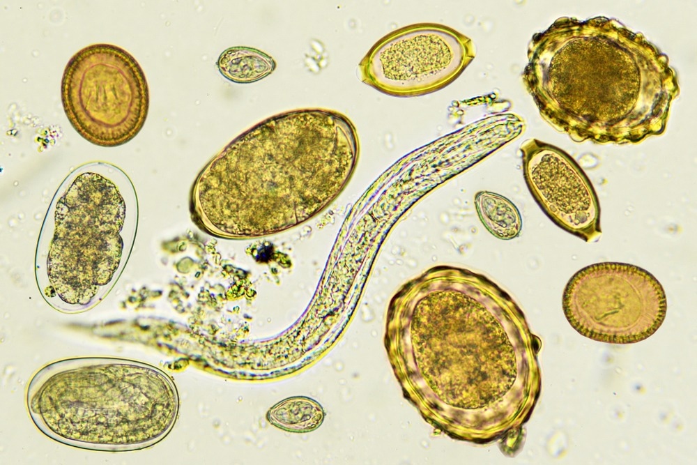
The impact of lung remodeling from prior helminth infection on SARS-CoV-2 disease pathogenesis
A recent study posted to the bioRxiv* preprint server reported that helminth exposure enhances viral clearance and survival of mice challenged with severe acute respiratory syndrome coronavirus 2 (SARS-CoV-2).

Background
Helminth endemic areas have reported lower coronavirus disease 2019 (COVID-19)-associated morbidity and mortality, raising speculations that helminth infections may modulate COVID-19 outcomes. A small study conducted in Ethiopia found a lower risk of severe COVID-19 in patients with helminth co-infection. However, the impact of helminth infections on pulmonary responses to SARS-CoV-2 remains unknown.
Prior studies suggest that helminth infections can be beneficial or may aggravate outcomes of viral infections. Indeed, previous infection, but not concurrent, with a lung-traversing worm was linked to beneficial outcomes after influenza challenge in mice. Moreover, individuals from helminth endemic regions have been likely exposed to worms in childhood.
The study and findings
In the present study, researchers evaluated the impact of lung remodeling from prior helminth infection on SARS-CoV-2 pathogenesis in mice. SARS-CoV-2-susceptible K18-hACE2 mice were infected with 500 larvae of Nippostrongylus brasiliensis and rested for 28 days to naturally clear the infection and recover. Subsequently, mice were challenged with a lethal dose of SARS-CoV-2 and observed for weight changes and survival.
Like worm-naïve mice (controls), worm-exposed animals rapidly lost weight upon the SARS-CoV-2 infection but showed a significantly higher survival rate (60%) than controls (20%). Lung viral loads were similar between controls and worm-exposed animals three days post-infection (dpi). However, N. brasiliensis-exposed mice had significantly reduced viral loads at 7 dpi.
Single-cell RNA sequencing (scRNA-seq) revealed that viral genes were less abundant in epithelial cells, endothelial cells, macrophages, neutrophils, and stromal cells in the lungs of worm-exposed mice. The lower viral titers at 7 dpi were suggestive of augmented adaptive responses instead of improved innate responses. Lymphocytic inflammation of lung parenchyma in worm-exposed mice was more pronounced than in controls.
The proportion of cluster of differentiation 8-positive (CD8+) T cells was markedly elevated in worm-exposed mice compared to controls at 7 dpi. SARS-CoV-2 spike-specific T cells increased in frequency by 7 dpi in worm-exposed mice. Depleting CD8+ T cells in worm-exposed mice before the SARS-CoV-2 challenge produced results similar to controls, suggesting that lung remodeling by prior helminth infection conferred protection against the viral challenge.
Further analysis indicated that the lungs of mice exposed to the helminth sustained the primed inflammatory state and long-term T helper type 2 (TH 2) cell signature. The researchers performed scRNA-seq on lung cells at 7 dpi and identified cell clusters. There was a loss of alveolar macrophages at 7 dpi, with cells expressing characteristic genes of alveolar macrophages accounting for a small proportion of the monocyte/macrophage compartment.
Two macrophage populations separated after re-clustering the monocyte/macrophage compartment and were differentially enriched in worm-exposed mice and controls, defined by the expression of Scgb1a1 (in controls) and Sec61a1 (in worm-exposed mice). The Scgb1a1 macrophages (from controls) showed elevated SARS-CoV-2 RNA levels, concordant with the increased lung viral loads in controls.
Genes involved in antigen processing/presentation were enriched in Sec61a1 macrophages from the worm-exposed mice, whereas those involved in inflammatory cytokine responses were enriched in Scgb1a1 macrophages. CD8+ T cells in the lungs of helminth-exposed mice secreted less pro-inflammatory cytokines such as tumor necrosis factor (TNF)-α and interferon (IFN)-γ. These cells had low expression of genes associated with inflammation and cytotoxic activity.
Spike-specific CD8+ T cells also produced less granzyme B in the lungs of helminth-exposed mice. The decreased T cell cytokine response in worm-exposed mice might result from lower viral loads or an increased regulatory response by CD4+ cells and macrophages. Finally, interstitial/alveolar macrophages were depleted with clodronate liposomes before the SARS-CoV-2 challenge.
Helminth-exposed mice depleted of alveolar macrophages no longer had significantly elevated CD8+ T cell responses. In contrast, the worm-exposed mice depleted of interstitial macrophages sustained high CD8+ T responses, suggesting that alveolar macrophages were required for superior CD8+ T cell responses. Nevertheless, interstitial macrophages were needed to control SARS-CoV-2 loads.
Conclusions
In summary, the study observed that prior N. brasiliensis infection of mice accelerated SARS-CoV-2 clearance and reduced mortality. This protection was mediated by the recruitment or expansion of SARS-CoV-2-specific CD8+ T cells. Moreover, helminth-primed pulmonary macrophages were necessary for the augmented viral clearance and T cell response.
The findings support the notion that lung-traversing helminths may reduce the severity of SARS-CoV-2 infection via macrophage-dependent enhancement of CD8+ T cells.
*Important notice
bioRxiv publishes preliminary scientific reports that are not peer-reviewed and, therefore, should not be regarded as conclusive, guide clinical practice/health-related behavior, or treated as established information.
- Hilligan, K. et al. (2022) "Helminth exposure protects against murine SARS-CoV-2 infection through macrophage dependent T cell activation". bioRxiv. doi: 10.1101/2022.11.09.515832. https://www.biorxiv.org/content/10.1101/2022.11.09.515832v1
Posted in: Medical Science News | Medical Research News | Disease/Infection News
Tags: Antigen, CD4, Cell, Coronavirus, Coronavirus Disease COVID-19, covid-19, Cytokine, Cytokines, Frequency, Genes, Helminths, Inflammation, Influenza, Interferon, Liposomes, Lungs, Macrophage, Monocyte, Mortality, Necrosis, Neutrophils, Respiratory, RNA, RNA Sequencing, SARS, SARS-CoV-2, Severe Acute Respiratory, Severe Acute Respiratory Syndrome, Syndrome, Tumor, Tumor Necrosis Factor

Written by
Tarun Sai Lomte
Tarun is a writer based in Hyderabad, India. He has a Master’s degree in Biotechnology from the University of Hyderabad and is enthusiastic about scientific research. He enjoys reading research papers and literature reviews and is passionate about writing.
Source: Read Full Article
