The Rebel Artist Who Revealed the Brain’s True Nature
Hypotheses come and go, but data remain. ― Santiago Ramón y Cajal

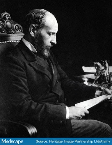
Santiago Ramon y Cajal (1852–1934).
From the vantage point of today ― a time when neurosurgeons regularly resect brain tissue to cure disease and rely on high-tech neuroimaging techniques to make diagnoses ― it’s hard to conceive of an era when artistic ability was as valuable to a neuroscientist as scientific knowledge and procedure.
But this was the case in the late 19th and early 20th centuries, when microscopy and photography — though established, maturing technologies — had yet to fuse to make microscopic images accessible to those outside the lab. As with astronomers gazing through telescopes in that bygone era, the accuracy of the images produced depended on who was doing the looking and, quite literally, how well they could draw.
When it came to late-19th century brain studies, things were complicated further by the nature of nervous tissue. Neurons were hard to see without special staining. Although scientists were getting used to the idea that the body was made of cells, they knew only of the smaller, more compact forms, such as those of blood cells and epithelial cells. The idea that cells could exist as long fibers with branches — what we now know as axons and dendrites — was simply not considered.
Except in the case of Santiago Ramón y Cajal (1852–1934), one of history’s most valuable contributors to scientific research and the understanding of brain anatomy and its cellular composition.
Readers may recall encountering him in the story of Rita Levi-Montalcini (1909–2012), whose home lab in Turin, Italy, was inspired by Ramón y Cajal’s in Valencia and who mastered his now famous silver nitrate staining method for nerve cells. But as we shall see, the silver nitrate method did not begin with Ramón y Cajal. Rather, he improved the method of Camillo Golgi (1843–1926), his Italian rival and the champion of a competing hypothesis, which held that the central nervous system (CNS) did not involve cells but instead an electrical reticulum. As Ramón y Cajal would prove, this idea was completely wrong. And yet, the two men would win the Nobel Prize together.
Coming of Age Alongside Cell Theory
Growing up in Petilla de Aragón, the young Ramón y Cajal was rebellious and occasionally mischievous, yet clever. At one point, he built a cannon that actually worked — so well that it destroyed the neighbor’s gate. Son of surgeon-anatomist Justo Ramón Casasús (1822–1903), the young Ramón y Cajal was not initially interested in medicine but in art and gymnastics. Throughout his life, he was taken with the works of Francisco de Goya (1746–1828), who also hailed from Aragon.
Nonetheless, he was nudged toward medicine by his father, who in 1869 took him to graveyards to collect and study bones. Th 16-year-old Ramón y Cajal began drawing their bony finds in intricate detail, which sparked his interest in anatomy and eventually led him to study medicine at the University of Zaragoza.
Excelling in anatomic dissection, he graduated in 1873, served briefly in the Spanish arm in Cuba, then returned to Zaragoza to earn a PhD, making him one of the earliest students to complete a physician-scientist program. This led to a series of academic appointments, beginning at Zaragoza and continuing through the universities of Valencia and Barcelona and finally to the Central University of Madrid.

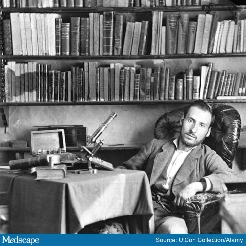
Santiago Ramon y Cajal as a student in 1876.
When Santiago Ramón y Cajal was born on May 1, 1852, the microscope was not new. Biologists had been viewing cells since 1665, when Robert Hooke (1635–1703) coined the term for the compartments he observed in a piece of cork, and cells remained a curiosity for quite a while. By the 1830s, however, Theodor Schwann (1810–1882) and Matthias Schleiden (1804–1881) had developed cell theory, the idea that the cell was the basic biological unit and that organisms and tissues were composed of cells.
In 1837, Czech anatomist Johann Purkinje (1787–1869) made thin slices through a cerebellum. Viewing the slices under a microscope, Purkinje visualized what we now know are neurons. But it was not a foregone conclusion that these fiber-shaped structures represented separate biological units comparable to the smaller, more spherical ones that researchers were recognizing as cells in blood and other tissues.
During a 6-decade career trajectory spanning these institutions, Ramón y Cajal would make discoveries throughout the body that encompassed phenomena such as muscle anatomy, inflammation, and microbiology, in a range of specimens, from insects to large animals, mammal embryos to bird brains. His improvement of the Golgi staining method would be the biggest contribution to Ramón y Cajal’s enduring legacy: the demonstration that the CNS was built of cells, just like the rest of the body.

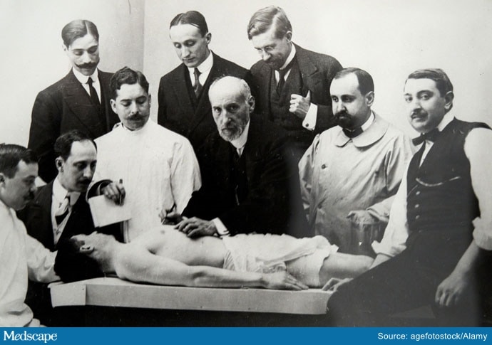
Dissection Class Given by Ramon y Cajal, 1915.
Working in Pavia, Italy, Golgi created the method in which nervous tissues were exposed first to potassium dichromate and subsequently to a silver nitrate solution. Published by Golgi the same year that Ramón y Cajal graduated from medical school, the technique led to the precipitation of silver chromate crystals, causing the cell membranes of neurons to appear black against a golden-yellow background. This was a major development, inasmuch as it revealed the nervous fibers that Purkinje had visualized, but in much more detail.
By 1887, Ramón y Cajal was not only trying the Golgi technique himself but was also deviating from it in radically creative ways. He adjusted the protocol, boosted concentrations of the agents, added methylene blue stain to the procedure, and cut thicker sections of nervous tissue than had been done previously.
Applying the tweaked technique to embryos and bird brains, Ramón y Cajal discovered that embryonic brain tissue lacked myelin, in contrast with adult brain tissue, and that as a consequence, embryonic and adult tissue differed in their capacity to hold stain. By using his improved stain, Ramón y Cajal also clearly saw that brains were composed of individual cells.
This finding supported the neuron hypothesis, or the Neuron Doctrine, the idea that the brain and spinal cord were composed of cells. That contradicted what Golgi had been proposing, the reticular hypothesis, or rather the reticular theory, for it actually had been the domain model of the CNS since the mid-19th century.
Essentially, the reticular theory saw the CNS as one big electrical network, which it is, but Golgi and his peers were thinking of a network consisting of an enormous web of conductive material, rather than stretched-out cells made of the same material as the cells they knew from viewing non-nervous tissues.

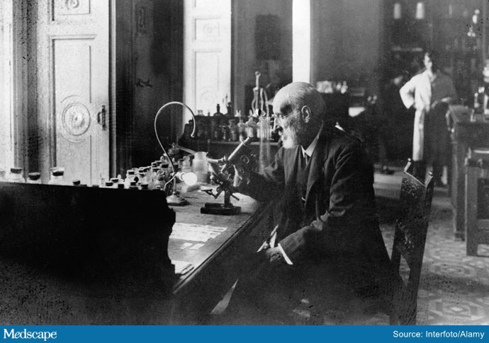
Ramon y Cajal at his microscope.
Viewing the CNS with Golgi’s stain, with or without Ramón y Cajal’s improvement, the tissue did have a reticular appearance. Throughout the 1890s, Ramón y Cajal harnessed his improved stain to hone in on the details of the cellular anatomy. He showed that there were cell bodies, axons, and dendrites ― in other words, that the various branchings were all parts of cells, albeit oddly shaped cells. In showing that the CNS was made of cells, Ramón y Cajal was also showing why it looked like a reticulum to Golgi and most of the neuroscience and anatomic community that agreed with him.
How ironic, and yet appropriate, that these two competing anatomists would end up sharing the 1906 Nobel Prize for their lifetimes of work, but also because of the technique that one invented and the other greatly enhanced, and in doing so, disproved one of their long-held theories on the brain.
David M. Warmflash, MD, is a freelance health and science writer living in Portland, Oregon. His recent book, Moon: An Illustrated History: From Ancient Myths to the Colonies of Tomorrow, tells the story of the Moon’s role in a plethora of historical events, from the origin of life, to early calendar systems, to the emergence of science and technology, to the dawn of the Space Age.
For more news, follow Medscape on Facebook, Twitter, Instagram, and YouTube.
Source: Read Full Article
