Tiny, ultra-flexible neural probes without cranial surgery open up new potentials for in vivo brain research

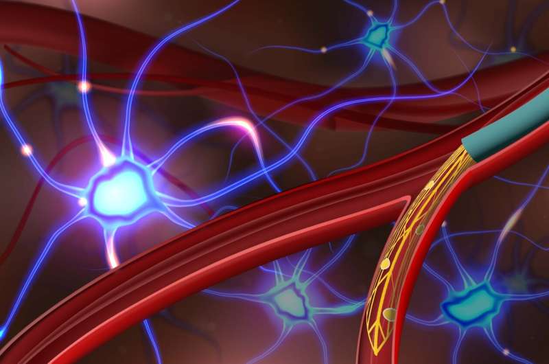
Researchers at Stanford University and Harvard Medical School have developed tiny and ultra-flexible mesh neural probes that can be implanted into sub-100-micrometer-scale blood vessels in the brains of rodents.
In their paper, “Ultraflexible endovascular probes for brain recording through micrometer-scale vasculature,” published in Science, the researchers demonstrate the potential of their device by measuring field potentials and single-unit spikes in the cortex and olfactory bulb of a rat without open skull surgery and without damaging the brain or vasculature. A Perspective piece in the same journal issue discusses the work done by the team.
The unique feature of this technology is the use of ultra-flexible endovascular probes that can be precisely delivered into tiny blood vessels without the need for invasive surgery. The probes can access brain regions that are difficult to reach safely with other methods, achieving selective implantation in different brain branches by tuning the mechanical properties of the probe.
Inspired by minimally invasive catheter-based injection procedures, the researchers designed polymer-based ultra-flexible micro-endovascular (MEV) probes that can be loaded into and injected from flexible microcatheters.
A saline flow through the microcatheter allows the probe to be then carried into deeper vasculature. The microcatheter is then retracted, leaving the MEV probes in place. Traditional intracranial depth electrodes for neuroelectronic interfaces require invasive surgery and can damage neural networks during implantation.
In histology testing, the probes demonstrated long-term stability with minimal immune response. Probes could not deform or penetrate the vessel walls, causing no damage to the blood-brain barrier, and did not significantly reduce blood flow or cause neurologic deficits.
In vivo electrophysiology recording was successfully achieved in the cortex and olfactory bulb of anesthetized rats. The probes demonstrated branch-selective implantation and operation, revealing different firing properties in neurological disease models. Single-unit activity recording was achieved, demonstrating single-cell resolution across vessel walls.
The cerebrovasculature ranges from large superficial cortical vessels to the microvasculature and capillary beds within the cortex. In the rat brain, ~5% of vessels have a diameter larger than 100 μm, which the study MEV probes could target.
Targeting smaller diameter vessels could be achieved by further reducing the size bending stiffness of the probes. The currently available endovascular probes for humans and sheep have only been able to target the largest vessel above 2.4-mm in diameter.
The study authors conclude that the “…platform technology could be extended to the detection and treatment of many neurological diseases as a research tool and could serve as the foundation for clinical translation of minimally invasive neuroelectronic interfaces.”
More information:
Anqi Zhang et al, Ultraflexible endovascular probes for brain recording through micrometer-scale vasculature, Science (2023). DOI: 10.1126/science.adh3916
Brian P. Timko, Neural implants without brain surgery, Science (2023). DOI: 10.1126/science.adi9330
Journal information:
Science
Source: Read Full Article
