meloxicam aman untuk ibu hamil

The outer layer of the brain, known as the cortex, is made of different types of neurons. Neuroscience studies suggest that these different neuron types have distinct functions, yet for a long time this was difficult to ascertain, due to the inability to examine and manipulate them in the brains of living beings.
In recent years, genetic techniques opened new possibilities for studying cells and their functions. Using some of these techniques, researchers at Forschungszentrum Jülich, effects of lorazepam with alcohol RWTH Aachen University, Cold Spring Harbor Laboratory and other institutes in the United States closely examined the functions of different pyramidal cells, which are commonly found in the human cortex.
Their findings, published in Nature Neuroscience, suggest that distinct types of pyramidal cells drive patterns of cortical activity associated with different brain functions. The team’s study builds on some of their previous works focusing on neuronal activity in the cortex.
“Our paper was based on one of our earlier studies, in which we used functional imaging to study the activity of the entire cortex of awake, task-performing mice,” Simon Musall, one of the researchers who carried out the study, told Medical Xpress.
“It was also based on a paper by our collaborators Katherine Matho and Josh Huang that introduced new transgenic mouse lines to target specific neural cell types. Putting the two together allowed us to selectively study the activity of highly specific neuron types throughout the entire cortex and ask whether their respective large-scale activity patterns are very distinct and functionally specialized or rather follow some general rules that might be defined by the function of known cortical areas.”
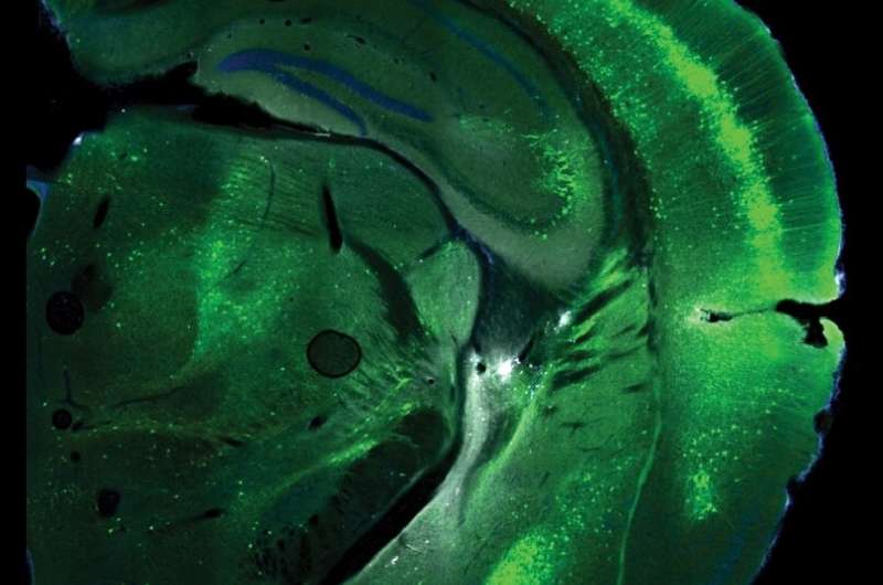
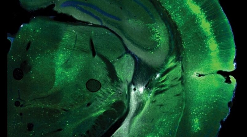
In their experiments, Musall and his colleagues first employed a microscopy technique called widefield calcium imaging to look at transgenic mouse lines expressing a fluorescent protein in a specific type of neuron throughout the cortex. When a cell becomes active, this protein emits a green light, allowing researchers to measure changes in this light that indicate when different parts of the brain become active.
Subsequently, the team measured the fluorescence in the mouse cortex. They did this using a highly sensitive camera system, measuring fluorescence light through the intact skull of awake mice in a non-invasive way.
“Most notably, we found that the cortex-wide activity patterns that we observed from different cell types were really distinct,” Musall explained. “This means that some brain regions were performing a specific function, such as processing sensory inputs, but this only became visible when measuring the activity of the specific cell type that carried this function.
“Conversely, many of these functions were not visible with non-specific imaging data because the activity of different cell types in the region got mixed. The same is true for most brain imaging tools that are routinely used in humans, such as fMRI, where we measure the activity of different brain areas based on oxygen consumption.”
The findings gathered by Musall and his colleagues delineate the functionally different cortical activity patterns that different types of pyramidal cells in the mouse brain elicit during decision-making. Even if all the cells seemed to play a key role in decision-making, different types exhibited different choice tuning patterns.
Overall, the researchers’ study highlights the value of new microscopy and genetic tools for unveiling the specific functions of distinct neuronal populations in the brain. In addition, it shows that the brain activity maps attained using conventional and unspecific imaging techniques only offer a limited view of the brain’s functional diversity.
In the future, this study could inspire new works aimed at examining the function of specific neuron populations in the cortex further. The researchers’ findings could also pave the way toward a better understanding of the complex neural underpinning of human decision-making.
“Each of these maps probably contains a myriad of far more detailed cell-type specific sub-maps that could reveal totally new activity patterns and brain functions that we are still unaware of,” Musall added. “So far, we have only studied the largest classes of neuron types, using the genetic tools that were available to us.
“We now plan to combine genetic and anatomical tools to gain even higher specificity and reveal at what point we obtain a more complete picture of large-scale activity patterns that capture the full functional range of different neuron subtypes.”
More information:
Simon Musall et al, Pyramidal cell types drive functionally distinct cortical activity patterns during decision-making, Nature Neuroscience (2023). DOI: 10.1038/s41593-022-01245-9
Journal information:
Nature Neuroscience
Source: Read Full Article
