urso luxury pens
In a recent study published in PNAS, researchers carried out real-time single-virion tracking in three dimensions to study the mechanisms and requirements of membrane attachment and fusion of severe acute respiratory syndrome coronavirus 2 (SARS-CoV-2) with the host cell.

Background
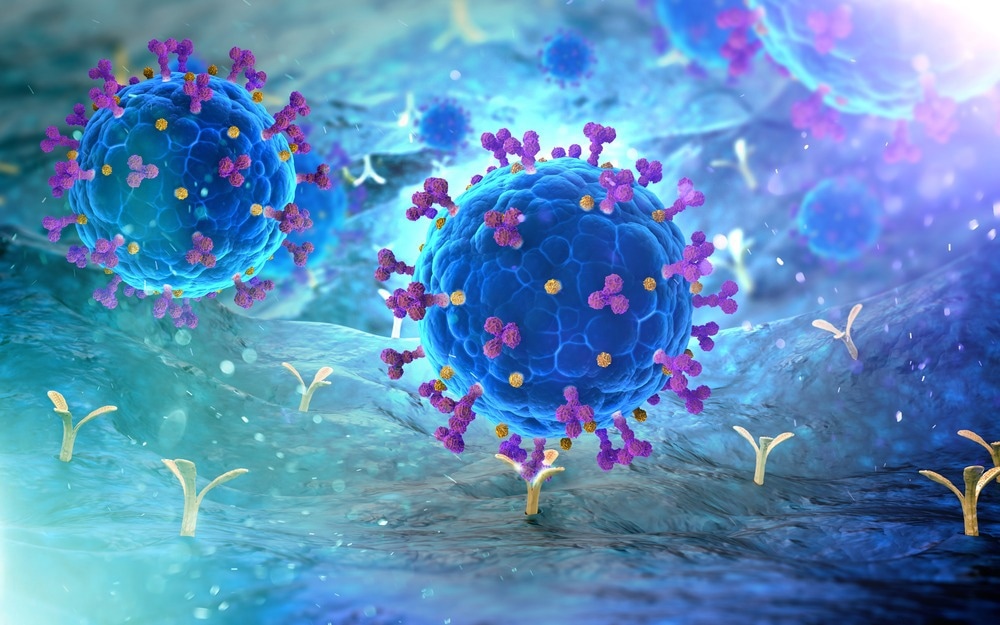
SARS-CoV-2 infection begins with the attachment of the viral spike protein with the angiotensin-converting enzyme 2 (ACE2) receptor on the host cell membrane. The viral entry into the cell can take two routes depending on the method of proteolysis of the spike protein.
The cleavage of the spike protein activates the viral fusion machinery. This cleavage can happen on the cell membrane surface through the action of two transmembrane serine proteases TMPRSS2 or TMPRSS4, following proteolytic activation by furin in producer cells, which results in noncovalently attached receptor binding subunit S1 and fusion subunit S2.
The cleavage can also occur through endosomal uptake and the action of endosomal cathepsins. Understanding the processing of the spike proteins and the action of specific proteases will help develop improved and targeted SARS-CoV-2 therapies.
About the study
In the present study, the researchers developed a novel method to directly visualize and live-track the host-cell membrane fusion and release of contents by a single virion in three dimensions. They used a chimeric virus consisting of vesicular stomatitis virus (VSV) with the endogenous glycoprotein gene (G) replaced with the spike protein from SARS-CoV-2. They also modified the structural phosphoprotein (P) of VSV replicative core to carry an enhanced green fluorescent protein (eGFP) to track the release of the virion contents into the cell.
The S protein is also labeled with a conjugated fluorescent dye, claritine dla dzieci opinie which will allow the S1 subunit release to be visualized in real-time as well. The VSV-eGFP-SARS-CoV-2 chimeras were made for the Wuhan -Hu-1 strain as well as Delta (B.1.617.2) and Omicron (B.1.1.529) strains. The infection assays with the VSV-eGFP-SARS-CoV-2 chimeras were carried out on cells grown in Dulbecco's Modified Eagle Medium (DMEM) with added HEPES (4-(2-hydroxyethyl)-1-piperazineethanesulfonic acid) and hydroxy dynasore to modulate the pH.
Real-time polymerase chain reaction (rt-PCR) with primers targeting SARS-CoV-2 ribonucleic acid (RNA)-dependent RNA polymerase was used to visualize the extent of viral replication. They also carried out similar assays with human isolates of SARS-CoV-2. Additionally, the role of endosomes in spike protein-mediated SARS-CoV-2 infection was explored using cells with a mutated gene for dynamin, which plays an important role in endocytosis.
Results
The study results uncovered a previously unknown requirement of a specific pH range of 6.5 – 6.8 for successful cell membrane fusion and cytosolic release of virion particles by the SARS-CoV-2 virion. This discovery also highlighted the role played by endosomes in the SARS-CoV-2 infection process. The fusion, cleavage, and release routes involving the serine proteases TMPRSS2 or TMPRSS4 were initially considered independent of endosomes. However, the requirement of an acidic pH indicates the involvement of endosomes in the release of virion contents, irrespective of the cleavage of the S1 subunit from the S2 subunit by the proteases.
The findings discuss three acidic pH-dependent routes of entry of the SARS-CoV-2 virus into the host cell. The virions with serine protease cleaved spike proteins undergo uptake and traffic to early endosomes, where the acidic pH of the early endosomes facilitates virion content release into the cytoplasm. The virions with the cathepsin cleaved spike proteins undergo uptake and cleavage in large endosomal compartments and release the contents into the cytosol of the host cell mediated by the low pH of the endosomal compartments.
A third route results in the release of virion contents at the cell surface if the membrane attachment occurs at a pH between 6.5 and 6.8. The study also found that the S1 subunit is shed from the spike protein during trimer formation with the ACE2 receptors at the cell membrane. This suggests an extended S2 fusion peptide intermediate at the host-cell membrane during neutral pH to keep the virion attached at the cell surface till endocytosis. The results also indicated that the spike protein on the Omicron variant was more resistant to proteolytic cleavage by furin.
Conclusions
The study results indicate pH-dependent viral tropism of SARS-CoV-2 virions in the respiratory mucosa. Cells in the nasal cavity expressing TMPRSS2 could facilitate rapid entry of SARS-CoV-2 since the pH of the nasal mucosa is between 6.2 and 6.8. Viruses that encounter the cells of the lung mucosa, which is at a more neutral pH, are likely to undergo uptake through endocytosis. The extended S2 intermediate at neutral pH offers a potential target for therapeutic action to combat SARS-CoV-2 infection.
- Kreutzberger, A. J. B., Sanyal, A., Saminathan, A., Bloyet, L.-M., Stumpf, S., Liu, Z., Ojha, R., Patjas, M. T., Geneid, A., Scanavachi, G., Doyle, C. A., Somerville, E., Correia, R. B. D. C., Di Caprio, G., Toppila-Salmi, S., Mäkitie, A., Kiessling, V., Vapalahti, O., Whelan, S. P. J., … Kirchhausen, T. (2022). SARS-CoV-2 requires acidic pH to infect cells. Proceedings of the National Academy of Sciences, 38. doi: https://doi.org/10.1073/pnas.2209514119 https://www.pnas.org/doi/10.1073/pnas.2209514119
Posted in: Medical Science News | Medical Research News | Disease/Infection News
Tags: ACE2, Angiotensin, Angiotensin-Converting Enzyme 2, Cell, Cell Membrane, Coronavirus, Coronavirus Disease COVID-19, Cytoplasm, Enzyme, Fluorescent Protein, Gene, Glycoprotein, Membrane, Omicron, pH, Polymerase, Polymerase Chain Reaction, Protein, Receptor, Respiratory, Ribonucleic Acid, RNA, SARS, SARS-CoV-2, Serine, Severe Acute Respiratory, Severe Acute Respiratory Syndrome, Spike Protein, Stomatitis, Syndrome, Virus
.jpg)
Written by
Dr. Chinta Sidharthan
Chinta Sidharthan is a writer based in Bangalore, India. Her academic background is in evolutionary biology and genetics, and she has extensive experience in scientific research, teaching, science writing, and herpetology. Chinta holds a Ph.D. in evolutionary biology from the Indian Institute of Science and is passionate about science education, writing, animals, wildlife, and conservation. For her doctoral research, she explored the origins and diversification of blindsnakes in India, as a part of which she did extensive fieldwork in the jungles of southern India. She has received the Canadian Governor General’s bronze medal and Bangalore University gold medal for academic excellence and published her research in high-impact journals.
Source: Read Full Article
