Biological Changes During Fatherhood
Testosterone changes in response to fatherhood
Neural changes that accompany fatherhood
Brain-level changes in the transition to fatherhood
Changes in prolactin in response to fatherhood
Neuro-endocrine changes caused by fatherhood
References
Further reading
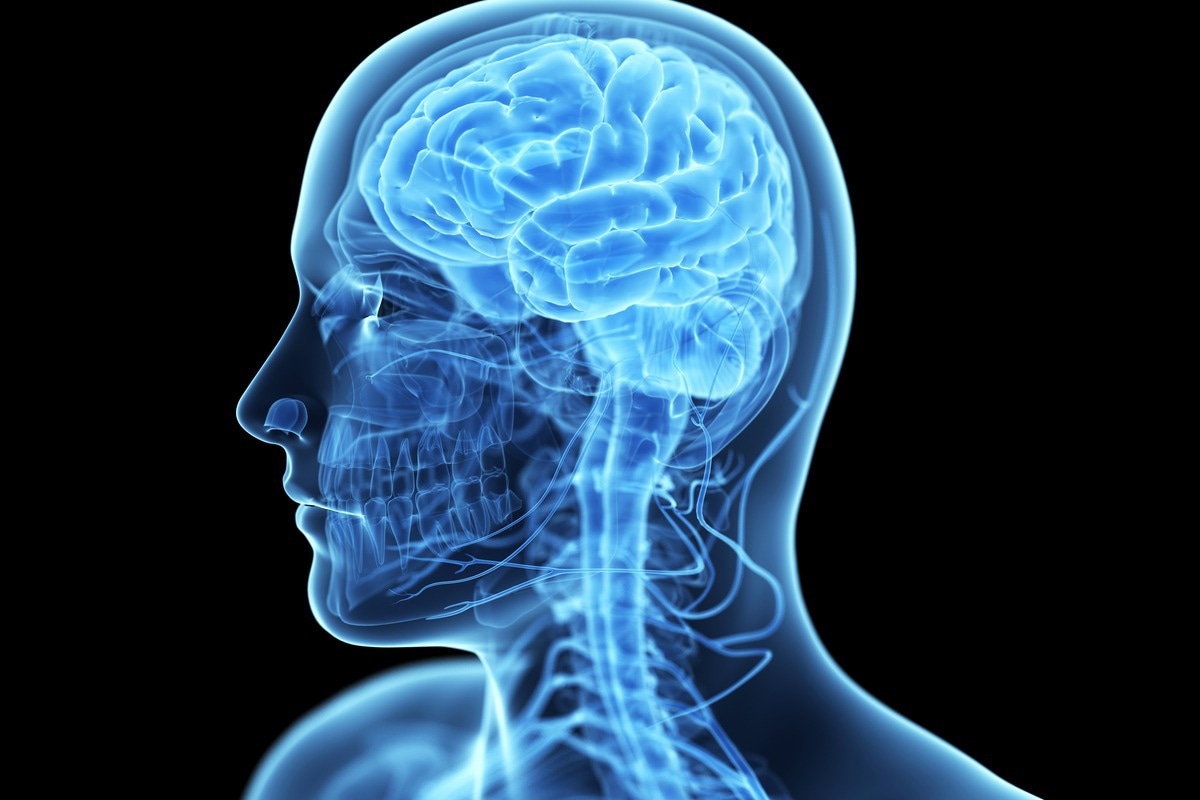
Humans undergo several neural and endocrine changes after the birth of their infant, which are thought to be associated with the caregiving responses exhibited by mothers and fathers. In particular, distinct and overlapping biological changes occur during fatherhood in response to exposure and close contact with that infant.

Image Credit: sofirinaja/Shutterstock.com
Testosterone changes in response to fatherhood
A pioneering study published in 2011 followed a representative group of 624 nonfather men in the Philippines between the ages of 21 and 26. Those who demonstrated high waking testosterone were more likely to become fathers at the follow-up time, 4.5 years later. In addition, those who became partnered fathers demonstrated large declines in waking and evening testosterone (an average of 34% when measured at night), significantly greater than the declines seen in single nonfathers.
This was consistent with the hypothesis that exposure to children (child interaction) is a testosterone suppressor. In this study, fathers who engaged in 3 or more hours of daily childcare had lower testosterone at follow-up compared to those who did not engage in childcare.
Consequently, researchers concluded that testosterone and reproductive strategy demonstrate bi-directional relationships; high testosterone predicates mating success but declines rapidly after fatherhood. Therefore, the study demonstrated a trade-off between mating and parenting – a phenomenon in this species that demonstrates father participation in childcare.
Neural changes that accompany fatherhood
Neural changes accompany fatherhood in man, and these changes are thought to be mediated by hormonal levels that start the exact change in paternal brain function. In a study that compared fathers of children aged 1–2 years relative to non-fathers, fathers demonstrated greater levels of plasma oxytocin and subsequent lower levels of plasma testosterone.
In addition, areas of the brain are important for face emotional processing (caudal middle frontal gyrus [MFG]), mentalizing (temporal-parietal junction [TPJ], Add regions involved in reward processing (medial orbitofrontal cortex [mOFC]) play the most strongly activated in fathers relative to non-fathers testosterone levels when negativity correlated with child stimuli and declined in inverse proportions to stimulation of the MFG.
Conversely, nonfathers demonstrated significantly stronger neural responses in the dorsal caudate and nucleus accumbens, regions of the brain involved in reward and approach-related motivation, when shown sexually provocative images.
Subsequently, these negative declines in testosterone foster a positive result concerning fatherhood at these transitions – namely, the augmented release of oxytocin and dopamine as a consequence of child interaction – are thought to be important to boost empathy towards children.

Image Credit: SciePro/Shutterstock.com
Brain-level changes in the transition to fatherhood
In a longitudinal study conducted in 2015, researchers investigated the structural changes in fathers' brains during the first four months postpartum. The research group examined 16 fathers to full-term, healthy infants whose brains were scanned using voxel-based morphometry and either 2–4 weeks postpartum or 12–16 weeks postpartum.
These fathers demonstrated increases in grey matter volume in positions that mapped to regions opening parental motivation from 2–4 weeks postpartum to 12–16 weeks postpartum. These include the hypothalamus, amygdala, striatum, and lateral prefrontal cortex.
However, grey matter in the orbitofrontal cortex, posterior cingulate cortex, and insula decreases. These patterns mimic the changes observed in mothers, with an overall increase in brain regions that would facilitate skills associated with parenting, such as nurturing, empathy, and the ability to perceive and react to baby behavior.
While activation in the brain regions is associated with empathy and the perception and understanding of child emotional behavior of common between mothers and fathers, a 2012 study demonstrated differences in parts of the brain that are activated for each parent. Mothers demonstrated higher activation in the amygdala region – and correlations between oxytocin release and the amygdala were found. By contrast, fathers demonstrated greater activation in circuits associated with social cognition and subsequently correlated with increased vasopressin.
The researchers, therefore, concluded that the motivational limbic activations between men and women might be gender-specific. The amygdala is associated with nurturing, care, and risk detection for mothers. In men, these regions are associated with cognitive functions such as thought, planning, problem-solving, and goal orientation. As such, the brains between fathers and mothers appear to have adapted in similar but convergent ways to enable dual care for infants. Both mothers and fathers have equal levels of motivation and are equally attuned to the needs of their child(ren).
Changes in prolactin in response to fatherhood
Prolactin, much like oxytocin, is an additional neuropeptide that can bind to trans membrane cytokine receptors in several tissues located on the periphery. These include the mammary glands and the central nervous system. Prolactin is conventionally associated with lactation and maternal behavior, and the facilitation of neurogenesis in mothers prompts maternal behavior.
While in mothers, prolactin levels increase during pregnancy and persist at the time of nursing, in fathers, particularly first-time fathers, prolactin levels dip approximately 30 minutes after exposure to their child. This effect has only been observed in first-time fathers, with subsequent exposure to infant cues, such as crying, causing prolactin levels to increase.
Therefore, fathers of subsequent children experience increased levels of prolactin. An initial drop in prolactin levels is associated with increased testosterone in first-time fathers. Speculation suggests that this may be an evolutionary mechanism that would prompt fathers to defend the child against any threatening stimuli that may have elicited infant cries.
Neuro-endocrine changes caused by fatherhood
Several works have compared differences in parents in response to infants' faces. Mothers tend to exhibit attentional bias and a higher preference for infants' faces than fathers. This persists in primary caregiving mothers compared to secondary caregiving fathers, who demonstrate greater activation of the brain areas associated with motivation and reward.
By contrast, primary caregiving fathers in same-sex relationships demonstrate increased functional connectivity with both the limbic – involved in behavioral and emotional response – as well as socio-cognitive networks – the cognitive processes related to the perception, understanding, and reaction to other people's behavior.
This has led researchers to suggest that parental differences may be shaped by more than just sex-specific biology. Instead, they are modulated by the type and amount of contact a parent has with their child following birth.
Studies have demonstrated that mothers and fathers can perceive infant emotions differently. Fathers tend to perceive happy emotions elicited by their infant less positively compared to mothers, which explains different behavior between the two genders, with subsequent knock-on effects on caregiving responses.
Research has demonstrated that fatherhood produces distinct biological changes to increase fathers' ability to care for their infants. There are distinct sex differences produced in the neuroendocrinology of men and women, which helps researchers understand how mothers and fathers contribute and react to parenting. It is evident from the literature that changes in exposure, most notably an increased involvement of fathers in the parental role, can be manipulated to optimize their infants' social, emotional, and developmental outcomes.
References
- Kim P, Rigo P, Mayes LC, Feldman R, Leckman JF, Swain JE. Neural plasticity in fathers of human infants. Soc Neurosci. 2014;9(5):522-535. doi:10.1080/17470919.2014.933713
- Mascaro JS, Hackett PD, Rilling JK. Differential neural responses to child and sexual stimuli in human fathers and non-fathers and their hormonal correlates. Psychoneuroendocrinology. 2014;46:153-163. doi:10.1016/j.psyneuen.2014.04.014
- Gettler LT, McDade TW, Feranil AB, Kuzawa CW. Longitudinal evidence that fatherhood decreases testosterone in human males. Proc Natl Acad Sci U S A. 2011;108(39):16194-9. doi: 10.1073/pnas.1105403108.
- Atzil S, Hendler T, Zagoory-Sharon O, Winetraub Y, Feldman R. Synchrony and specificity in the maternal and the paternal brain: relations to oxytocin and vasopressin. J Am Acad Child Adolesc Psychiatry. 2012:798-811. doi: 10.1016/j.jaac.2012.06.008.
- Grebe NM, Sarafin RE, Strenth CR, Zilioli S. Pair-bonding, fatherhood, and the role of testosterone: A meta-analytic review. Neurosci Biobehav Rev. 2019;98:221-233. doi: 10.1016/j.neubiorev.2019.01.010.
- Jones BC, Hahn AC, Fisher CI, Wang H, Kandrik M, Han C, Fasolt V, Morrison D, Lee AJ, Holzleitner IJ, O'Shea KJ, Roberts SC, Little AC, DeBruine LM. No Compelling Evidence that Preferences for Facial Masculinity Track Changes in Women's Hormonal Status. Psychol Sci. 2018;29(6):996-1005. doi: 10.1177/0956797618760197
Further Reading
- All Parenting Content
- What is Helicopter Parenting and Why is it Bad?
Last Updated: Jul 4, 2022

Written by
Hidaya Aliouche
Hidaya is a science communications enthusiast who has recently graduated and is embarking on a career in the science and medical copywriting. She has a B.Sc. in Biochemistry from The University of Manchester. She is passionate about writing and is particularly interested in microbiology, immunology, and biochemistry.
Source: Read Full Article
