What are Antinuclear Antibodies?
Antinuclear antibodies (ANA) are autoantibodies directed against the nuclei of some of one’s own body cells. They are present in the blood in persons with certain autoimmune disorders.
An autoimmune condition is one which results in the immune system of an individual fighting the cells and tissues of the same individual rather than those which are from a foreign organism. The disorder can be both serious and chronic.
Antibodies are large protein molecules formed by the immune system to tag certain specific molecules (called ‘antigens’) on foreign or non-self substances, so that immune cells can latch on to them and destroy them. When these are formed against normal body cells instead, they are called autoantibodies. ANAs are autoantibodies directed against certain antigens on the nuclei of the body’s own cells.

Skip to
- What is ANA testing used for?
- Indications for ANA testing
- How is the ANA test performed?
- What do ANA results mean?
What is ANA testing used for?
Testing for ANA is a commonly used serological test for autoimmune disease.
ANA testing was first carried out by Holborow as long back as 1957, using an indirect immunofluorescence technique. ANA tests help to diagnose autoimmune disorders like:
- Rheumatoid arthritis in which there is pain and swelling of the joints of the hands and the feet, mostly, though other joints are often affected
- Systemic lupus erythematosus or SLE occurs in a chronic inflammation of the joints, kidneys, blood vessels, and brain
- Sjogren’s syndrome in which the patient typically experiences dryness of the eyes and/or mouth, because of a chronic destructive inflammation of the parotid salivary gland and the lacrimal (tear) gland of the eyes
Indications for ANA testing
There are certain symptoms which indicate a high probability of lupus or other autoimmune disorders, such as fever, joint pain and swelling, rashes (which are butterfly-shaped on the face in lupus), muscle pain and tiredness.
How is the ANA test performed?
A blood sample is obtained and checked for the presence of ANA using the indirect immunofluorescence (IF) or enzyme-linked immunosorbent assay (ELISA).
Both are based upon incubating the patient serum with nuclear antigens in suspension that have been coated on wells in a microplate. Following incubation, excessive antibodies are removed by washing.
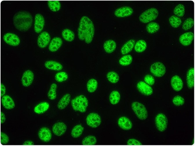
In IF-ANA the bound antibodies are detected using another antibody which binds to human antibody molecules, and which carries a fluorescent molecule. This means that the presence of ANA will show up as fluorescence on the cells when examined using a fluorescent microscope.
In ELISA, the antigen-antibody complexes are detected by another antibody which binds to human antibodies, but which carries an enzyme as a marker. When ANA is present in the sample, the enzyme will remain in the wells, which contain the complexes, and a substrate that is then added will change color as a result of enzyme activity.
What do ANA results mean?
A positive ANA test means that the antibodies were detected in the individual’s blood sample. This may indicate:
- The presence of SLE
- The presence of another autoimmune disorder, though not all autoimmune disorders are associated with ANA
- A viral infection
- Chronic infection
- Some malignancies
- Autoimmune thyroid disease
Sometimes, ANA testing turns out positive in a healthy person, or in some cases, following the ingestion of certain medications. In fact, after the age of 65 years, about one in three healthy adults tend to have ANA in their blood.
In a random screening, about 5% will have high enough levels of ANA to be considered positive. In fact, for every individual with SLE, there would be about 50 people without autoimmune disease but with a positive ANA. This means that ANA testing should not be used alone to diagnose any disease. Clinical criteria must be satisfied as well. However, in some patients the first presentation is a positive ANA test, to be followed by clinical features after a few years.
For this reason an ANA test is considered positive only if the level is significantly higher than the normal concentration. The report contains the intensity of reaction (corresponding to the ANA quantity) as well as the staining pattern, which reflects the way the antibody binds to the nucleus. This shows which antigens are being bound and offers clues as to which type of autoimmune disorder may be present.
The ANA titer is reported in terms of the dilution at which the test remains positive. Another method is reporting the intensity of fluorescence in international units per ml (IU/ml) at a dilution already set. In most cases, the limit for positivity is typically >7 IU/ml.
Some significant staining patterns include:
- Homogeneous staining in SLE or mixed connective tissue disease
- Speckled pattern in SLE and Sjogren’s syndrome because the antibodies target the nucleosomes, units which form the fundamental structure of coiled compacted chromatin
- Nucleolar pattern in diffuse systemic sclerosis
- Centromere staining in limited scleroderma
- Peripheral or rim staining in SLE or scleroderma
Sources
- https://medlineplus.gov/lab-tests/ana-antinuclear-antibody-test/
- https://www.racgp.org.au/afp/2013/october/antinuclear-antibody-test/
- https://www.ncbi.nlm.nih.gov/pubmed/15041045
- https://academic.oup.com/rheumatology/article/39/6/581/1783869
- cvi.asm.org/…/article-info?versioned=true
Further Reading
- All Antibody Content
- Antibody – What is an Antibody?
- Antibody Forms
- Antibodies in Medicine
- Antibody Structure
Last Updated: May 9, 2019

Written by
Dr. Liji Thomas
Dr. Liji Thomas is an OB-GYN, who graduated from the Government Medical College, University of Calicut, Kerala, in 2001. Liji practiced as a full-time consultant in obstetrics/gynecology in a private hospital for a few years following her graduation. She has counseled hundreds of patients facing issues from pregnancy-related problems and infertility, and has been in charge of over 2,000 deliveries, striving always to achieve a normal delivery rather than operative.
Source: Read Full Article
