
allied long reach
Single molecule localization microscopy (SMLM) is a group of super-resolution microscopy techniques that can obtain images at extremely high resolutions. These highly advanced tools have been used to aid discoveries within the biomedical field.
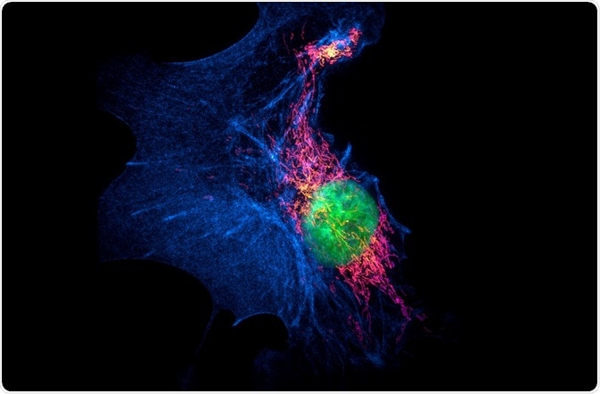
MichaWeber | Shutterstock
What is Single-Molecule Localization Microscopy (SMLM)?
SMLM uses the stochastic super-resolution method that relies on the complex behavior of molecular light sources to make fluorophores emit light at separate times, allowing different parts of a sample to be imaged individually.
The common types of SMLM include:
- Photo-activated localization microscopy (PALM)
- Fluorescence photo-activation localization microscopy (fPALM)
- Stochastic optical reconstruction microscopy (STORM)
- Cryogenic optical localization in 3D(COLD)
Types of Single-Molecule Localization Microscopy
PALM and fPALM
Both PALM and fPALM involve collecting many images under a microscope, each with just a few active isolated fluorophores. The fluorophores are activated from a less-emissive state into a brighter one and then back to the less-emissive state.
The position of the fluorophores is calculated with great precision and this information is built up into a single image. Both PALM and fPALM use fluorophores instead of photo-switchable dyes. The difference lies in the microscope, as PALM uses a total internal reflection fluorescence (TIRF) microscope and fPALM uses a confocal microscope.
STORM
STORM uses two lasers and photo-switchable dyes in combination with TIRF microscopy. The dye is attached to the protein of interest via immunohistochemistry. One laser turns the fluorophores on and the other turns them off. Multiple separate images of the sample are then processed and merged into one final image. The time resolution is faster compared to PALM, as it activates and images the fluorescence simultaneously rather than in cycles.
COLD
COLD localizes many fluorescent sites within a small to medium sized biomolecule with angstrom-scale resolution. This technique uses low temperatures which allows for more photons to be emitted by the sample before it is damaged by photobleaching. The 2D projections gained from the fluorophores can also be reconstructed into a 3D image.
Applications of SMLM
Localization microscopy can be applied to single fluorescent molecules without the need for a photo-conversion process. This saves time and allows a wide range of molecules to be examined.
PALM is very useful for single particle tracking, as very small number of fluorophores are excited at any time which gives them no diffraction overlap.
COLD imaging can be used to visualize the proteins in mitochondrial and nuclear membranes. It has also been used to visualize the estrogen receptor Her2, an indicator of breast cancer.
SMLM techniques have been used to image:
- The cell membrane
- G-protein coupled receptors (GPCR)
- Exosomes of human breast and cervical cancer cells
- Tracking N- and O-linked glycans on the membrane of live mammary cancer cells
- The organization of proteins on the plasma membrane of cells
Perspectives & conclusions
The time resolution of SMLM techniques can be improved which limits their use in very fast biological processes. Microscopy techniques can produce a lot of noise that interferes with the imaging and SMLM are no exception to this.
Improving this signal-to-noise ratio will benefit these imaging techniques. To conclude, SMLM techniques, alone and in combination with other methods will definitely provide more information on a wide variety of biological processes in the future.
Sources
- https://www.sciencedirect.com/science/article/pii/S0006349506721403
- https://www.nature.com/articles/nmeth929
- https://www.nature.com/articles/nmeth.4141
- https://www.ncbi.nlm.nih.gov/pmc/articles/PMC5520217/
- bitesizebio.com/…/
Further Reading
- All Super-resolution microscopy Content
- Stochastic Optical Reconstruction Microscopy (STORM)
- Structured Illumination Microscopy (SIM)
- Stimulated Emission Depletion (STED) Microscopy
- Confocal Versus Super-resolution Microscopy
Last Updated: Dec 20, cheap seroquel coupon no prescription 2018
Written by
Samuel Mckenzie
Sam graduated from the University of Manchester with a B.Sc. (Hons) in Biomedical Sciences. He has experience in a wide range of life science topics, including; Biochemistry, Molecular Biology, Anatomy and Physiology, Developmental Biology, Cell Biology, Immunology, Neurology and Genetics.
Source: Read Full Article