buy online valtrex canadian pharmacy no prescription
Lungs are complex organs that are often the site of disease or infection. Studying lungs can be difficult, due to their complicated structure, but lung organoids can aid in this. Lung organoids are collections of lung-derived epithelial cells grown in a 3D culture, without the structural supporting cells they grow within the lung. In this setting, the cells form the self-forming structures referred to as “lung organoids”.

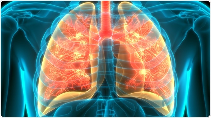
Image Credit: Magic mine/Shutterstock.com
How lung organoids are made
Lungs are composed of complex branching systems, lined with highly specialized cells. These cells facilitate several crucial functions, compazine side effects such as oxygen exchange, but also immune functions to protect this vulnerable part of the body from infection. This makes them an important area of study in current medical research. However, studying cells is often done with cell cultures, which are not always adequate for understanding complex interactions.
Cell cultures have largely been grown in a monolayer which lacks more complex in vivo interactions between cells and cell types. Growing cells in 3D organoids offer a more complex and realistic insight because organoids fulfill three important aspects of in vivo organs: more than one cell type can be grown together, these cell types can structure themselves approximately like they do in the native organ and retain some innate functioning.
The production of organoids relies on the same processes that take place during organ production in normal development. These processes include specific adhesion properties which give rise to the self-organization of cells and differentiation of progenitor cells which is limited to certain areas. This can be done using embryonic tissue progenitor cells, which will organize into structures characteristic of early organogenesis. Mature cells from adult organ tissue can also be grown into organoids but to a lesser extent.
Human lung organoids have been developed from pluripotent stem cells, which can be transplanted into mice where growth continues into the airways and alveoli of lungs. The result after 6 months of culturing can be equivalent to the human lungs in the second trimester of gestation. A similar result could be achieved when the lung bud organoids remained in culture.
Research and applications
Organoids provide a realistic, animal testing-free method to study tissue and organ development. One of the most common applications of lung organoids is to study how cells respond to damage.
Normally, basal cells in the lung, which are part of the epithelial cells, change behavior and proliferation in response to cell damage to restore the epithelial layer but can also become stem cells in response to direct damage. Lung organoids can be used to elucidate more about the regeneration mechanisms of the epithelial barrier and to screen for drugs regulating this process.
Infections can also be studied using lung organoids. Respiratory syncytial virus (RSV) affects newborns and currently has no licensed effective treatment. There are no models capable of reproducing RSV infection.
However, when lung organoids were infected with RSV, the epithelial cells of the organoid were shed into the lumen of the branching structures in a manner previously seen and in concurrence with symptoms the virus causes. This presents lung organoids as viable models to study difficult pulmonary diseases, such as RSV.
In addition to strictly viral infections, the potential to use lung organoids in genetic conditions has also been investigated. Hermansky-Pudlak Syndrome (HPS) is, in some forms, associated with pulmonary fibrosis. When HSP-associated pulmonary fibrosis was partially induced in lung organoids, there were less sharp branching structures and accumulation of mesenchymal cells.
This experiment also found support for the theory that certain forms of pulmonary fibrosis are caused by injury to epithelial tissue. Therefore, it seems possible that lung organoids are so functionally similar to lungs that it can be possible to model some forms of pulmonary fibrosis using them.
Limitations of lung organoids
Several challenges still face lung organoids and their use in research. As mentioned, a lung organoid matching the developmental stage of the second trimester was grown in 6 months. This indicates that organoid maturation matches the pace of normal development. However, reaching the full maturation stage remains a challenge.
Another current limitation is that the branching process that takes place appears to be random. While this in congruence with a theory stating the branching process will take place in a space-filling manner, this theory is not proven and the randomness can present potential difficulties.
Lastly, the exact mechanism of the patterning and general nature of the organoid mesenchymal cells is unknown. However, lung organoids still hold much potential as models for lung functioning, disease, and drug screening purposes.
Sources
- Barkauskas, C., Chung, M., Fioret, B., Gao, X., Katsura, H., and Hogan, B., 2017. Lung organoids: current uses and future promise. Development, 144(6), pp. 986-997.
- Chen, Y., Huang, S., de Carvalho, A., Ho, S., Islam, M., Volpi, S., Notarangelo, L., Ciancanelli, M., Casanova, J., Bhattacharya, J., Liang, A., Palermo, L., Porotto, M., Moscona, A. and Snoeck, H., 2017. A three-dimensional model of human lung development and disease from pluripotent stem cells. Nature Cell Biology, 19(5), pp. 542-549.
- Nadkarni, R., Abed, S., and Draper, J., 2016. Organoids as a model system for studying human lung development and disease. Biochemical and Biophysical Research Communications, 473(3), pp. 675-682.
Last Updated: Sep 24, 2020

Written by
Sara Ryding
Sara is a passionate life sciences writer who specializes in zoology and ornithology. She is currently completing a Ph.D. at Deakin University in Australia which focuses on how the beaks of birds change with global warming.
Source: Read Full Article
