codeine phosphate syrup manufacturer
 *Important notice: medRxiv publishes preliminary scientific reports that are not peer-reviewed and, therefore, should not be regarded as conclusive, effet secondaire avec champix guide clinical practice/health-related behavior, or treated as established information.
In a recent study posted to the medRxiv* preprint server, researchers observe persistent severe acute respiratory syndrome coronavirus 2 (SARS-CoV-2) replication in severe coronavirus disease 2019 (COVID-19) patient lungs.
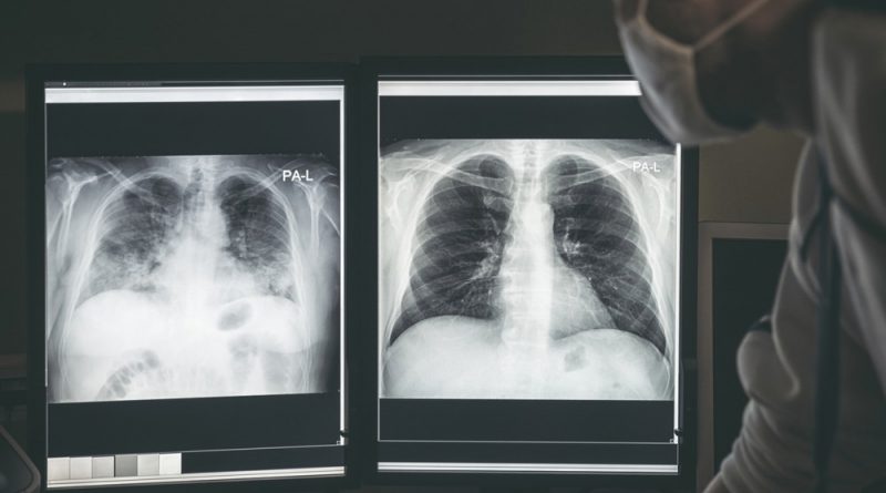
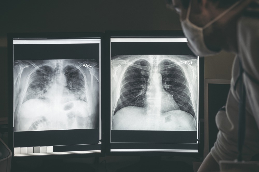 Study: SARS-CoV-2 Viral Replication Persists in the Human Lung for Several Weeks after Onset of Symptomatic Severe COVID-19 and Is Associated with Attenuated Pulmonary Immunity and Variant-Specific Clinical Sequalae. Image Credit: Jaroslav Moravcik / Shutterstock
Persistence of SARS-CoV-2 during severe COVID-19
COVID-19 has significantly contributed to global mortality. Notwithstanding recent research advances, severe COVID-19 pathogenesis with respect to viral kinetics remains poorly understood.
Postmortem studies have reported persistent virus in tissues by immunohistochemistry or polymerase chain reaction (PCR) assay. SARS-CoV-2 detected in these analyses may not be replication-competent, which can only be determined by viral culture.
Further, some studies have demonstrated the shedding of replicative virus from the upper airways that last up to eight days following symptom onset. Nevertheless, information about the persistence of replicative virus in the lower airways is scarce, especially among patients with severe COVID-19 requiring mechanical ventilation.
About the study
In the present study, researchers determine whether SARS-CoV-2 replication in severe COVID-19 persists beyond 10 days in the lower respiratory tract, independent of viral persistence in the upper airways.
To this end, the researchers ascertained the duration and frequency of replication-competent virus in the lungs. Lung samples were obtained through a needle biopsy within two hours of death from 42 mechanically ventilated patients during the Beta and Delta waves.
Study findings
Culturable virus was detected in 38% of patients for up to four weeks after symptom onset. Contrastingly, in a control cohort of ambulatory subjects, culturable virus was detected in one nasopharyngeal swab sample at day 12 post-onset but not in samples collected on day 19. Notably, multiple organs were positive for SARS-CoV-2 by PCR in Delta cohort decedents who were positive for the culturable virus.
Clinical characteristics were similar between culture-positive and -negative groups. The cycle threshold (Ct) value of nasopharyngeal swab samples at admission and death was not associated with culture positivity.
Only culture-positive Delta cohort subjects had a shorter interval between symptom onset and death, more secondary bacterial infections, and increased rates of concurrent bronchopneumonia than culture-negative individuals.
Omics eBook

Immunohistochemical staining revealed a significantly reduced infiltration of cluster of differentiation 4-positive (CD4+) macrophages and T-cells, as well as CD8+ T-cells, in the interstitial and alveoli of culture-positive subjects in the Delta cohort relative to culture-negative decedents. Immunohistochemical analysis was not performed for Beta cohort decedents.
Similarly, an attenuated CD4+ and CD8+ T helper type 1 (TH1) cell response was observed in cells from the deconstructed lung tissues of decedents from the Delta cohort.
The researchers also performed a transcriptional analysis of the lungs and identified 630 upregulated and 885 downregulated genes between culture-positive and -negative individuals.
The upregulated genes were involved in pathways associated with pro-inflammatory responses, whereas downregulated genes were primarily associated with homeostasis. After adjusting for multiple factors, 11 upregulated and five downregulated genes were identified in culture-positive individuals. Patterns of T-cell exhaustion were not evident.
Finally, the team assessed whether any differentially expressed gene could be a biomarker distinguishing culture-positive individuals from culture-negative subjects. Logistic regression identified gremlin-1 (GREM1) and fibroblast growth factor-binding protein 1 (FGFBP1) with high sensitivity and specificity.
Conclusions
Taken together, the study findings demonstrate the simultaneous occurrence of viremia and hyperinflammation in the third and fourth weeks of COVID-19, which contradicts the view that these phases occur successively. The pro-inflammatory and antiviral responses were over-represented in culture-positive decedents.
The researchers did not observe attenuated type 1 interferon (IFN) responses. The typical histologic characteristics of severe COVID-19, including microvascular thrombosis and diffuse alveolar damage, were comparable between culture-positive and -negative groups, thus implying that they occurred in the early phase of the disease.
Notably, culture-negative decedents exhibited higher pneumocyte infiltration than culture-positive subjects. These results are relevant to patients with severe COVID-19 and acute respiratory distress syndrome (ARDS) and likely inapplicable to patients with mild disease. Nevertheless, only patients during the Beta- and Delta-predominant periods were considered; therefore, these findings do not necessarily apply to infection with the SARS-CoV-2 Omicron and its subvariants.
A control cohort of subjects with ARDS of other etiologies was also lacking. Overall, the study findings have potential implications for using antivirals in critically ill patients.
Nevertheless, there remains an urgent need to identify better biomarkers for phenotypes and patient subsets, which may benefit from the concurrent use of antiviral and anti-inflammatory therapies.
*Important notice: medRxiv publishes preliminary scientific reports that are not peer-reviewed and, therefore, should not be regarded as conclusive, guide clinical practice/health-related behavior, or treated as established information.
- Preliminary scientific report. Tomasicchio, M., Jaumdally, S., Pooran, A., et al. (2023). SARS-CoV-2 Viral Replication Persists in the Human Lung for Several Weeks after Onset of Symptomatic Severe COVID-19 and Is Associated with Attenuated Pulmonary Immunity and Variant-Specific Clinical Sequalae. medRxiv. doi:10.1101/2023.03.06.23286834. https://www.medrxiv.org/content/10.1101/2023.03.06.23286834v1.
Posted in: Medical Science News | Medical Research News | Medical Condition News | Disease/Infection News | Healthcare News
Tags: Acute Respiratory Distress Syndrome, Anti-Inflammatory, Assay, Biomarker, Biopsy, CD4, Cell, Coronavirus, Coronavirus Disease COVID-19, CT, Exhaustion, Fibroblast, Frequency, Gene, Genes, Growth Factor, immunity, Immunohistochemistry, Interferon, Lungs, Mortality, Nasopharyngeal, Omicron, Phenotype, Polymerase, Polymerase Chain Reaction, Protein, Research, Respiratory, SARS, SARS-CoV-2, Severe Acute Respiratory, Severe Acute Respiratory Syndrome, Syndrome, T-Cell, Thrombosis, Virus

Written by
Tarun Sai Lomte
Tarun is a writer based in Hyderabad, India. He has a Master’s degree in Biotechnology from the University of Hyderabad and is enthusiastic about scientific research. He enjoys reading research papers and literature reviews and is passionate about writing.
Source: Read Full Article