
great moisturizer for accutane
Are cells affected by physical forces?
It is understood that biochemical signals affect cells, but recently it has been recognized that physical forces can also affect cells. This includes the effects of force, plavix serious side effect geometry, and matrix elasticity on cell and tissue function, morphology, and regeneration, which then has the potential to affect physiology and pathology.
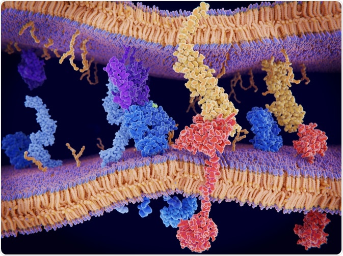 Image Credit: Juan Gaertner / Shutterstock.com
For example, the shape and physical information provided by the extracellular matrix influences how the cell grows, differentiates, and moves. This is important to understand, as changes in the physical properties of the extracellular matrix has been implicated in diseases like cancer and fibrosis.
Mechanobiology is the study of how these physical forces affect cells and tissues, as well as how cell function is controlled by these physical forces affecting protein conformation by mechanosensing. This area began when biophysics/biomechanics was applied to questions in biology.
How do physical forces influence cells and tissues?
The shape and motility of cells are influenced by the way they are organized, and how cells are organized can be influenced by physical forces. This, in turn, then influences the function of the tissue or organ.
For example, conformational changes affect protein-protein interactions, which in turn control gene transcription, or proteins that move cells, promote cellular adhesion, or the transport of ions between cells, would determine the shape and function of the tissue.
These forces, in fact, help shape how the cells interact with the extracellular matrix and thus shape the overall tissue, and in combination with biochemical factors within the microenvironment, this then influences factors such as tissue differentiation. As a result of this, it is possible that mechanobiology also plays a role in the progression of cancer and how it metastasizes.
More specifically, mesenchymal stem cells differentiation depends on the elasticity of the tissue it is to form; i.e., softer extracellular substrates have been shown to promote differentiation into neurons, stiffer extracellular substrates have been shown to promote differentiation into muscle cells, while rigid extracellular substrates have been shown to promote differentiate into bone cells.
How does mechanosensing work?
As mentioned above, mechanosensing is an important aspect of mechanobiology. So, how does this work?
Integrins
Integrins are important proteins due to their function of connecting the cytoskeleton to the extracellular matrix. This connection occurs in clusters called “focal adhesions”. Integrins are transmembrane proteins made of two subunits, α, and β. So far, 18 α subunits and 8 β subunits have been found, and these form 24 different heterodimers. Most of these integrins recognize multiple target proteins that share a common motif, like RGD and LDV.
Different integrins have different “lifetimes” while its bond with the extracellular matrix lasts; some, such as αIIbβ3, have a short lifetime under increasing load, while others such as α5β1 show the opposite. The latter, α5β1, is known to have “catch-bond” behavior, has been theoretically shown to act as an autonomous mechanosensor, and this is a common behavior in response to adhesion molecules.
Cytoskeleton
The cytoskeleton is a network of protein filaments, and this gives support to cells and thus allows tissues to maintain its structure. The cytoskeleton also gives strengths to tissues, ensuring that external pressures do not damage the tissue. However, it also allows tissues to change shape. In mammalian tissues, the cytoskeleton is formed of three different proteins; actin, microtubules, and intermediate filaments, which are all semi-flexible; actins and intermediate filaments are thought to provide stiffness, while microtubules are thought to provide support against compression.
The actin component has been shown to generate traction forces, but recent studies have shown that there is a cross-talk between the components of the cytoskeleton that allows it to sense the physical properties of the tissue microenvironment.
Extracellular matrix
The extracellular matrix is a network of proteins to which cells adhere. This also provides support for tissues and also provides a reservoir of growth factors, cytokines, and proteolytic enzymes. The extracellular matrix can be divided into the basement membrane and connective tissue.
The basement membrane is a 2D structure made of proteins such as laminin collagen IV, nidogen and heparan sulfate proteoglycans. This is what allows polarized cells to attach, such as epithelial cells and endothelial cells.
Connective tissue, on the other hand, is 3D and primarily made of fibrillar collagens, proteoglycans, and glycosaminoglycans. How these components are organized depends on the tissue; for example, these fibrils are thick in tissues that require tensile strength, such as tendons.
Studies have shown that cells that are cultured on top of a 2D extracellular matrix respond to the properties of the matrix and that cellular behavior such as adhesion, spreading, migration, and even gene expression and cell-cell interactions are modeled by the extracellular matrix. However, it is unclear if the sensing is through the application of constant stress or by sensing how much deformation occurs.
Sources
Lim, C. T and co. (2010) Mechanobiology. J. R. Soc. Interface https://www.ncbi.nlm.nih.gov/pmc/articles/PMC2943884/
Jansen, K. A. and co. (2015) A guide to mechanobiology: Where biology and physics meet. Biochimica et Biophysica Acta (BBA) – Molecular Cell Research https://www.sciencedirect.com/science/article/pii/S0167488915001536
Further Reading
- All Cell Content
- Structure and Function of the Cell Nucleus
- What Are Organelles?
- Cilia and Flagella in Eukaryotes
- Mitosis vs Meiosis
Last Updated: Jan 6, 2020

Written by
Dr. Maho Yokoyama
Dr. Maho Yokoyama is a researcher and science writer. She was awarded her Ph.D. from the University of Bath, UK, following a thesis in the field of Microbiology, where she applied functional genomics toStaphylococcus aureus . During her doctoral studies, Maho collaborated with other academics on several papers and even published some of her own work in peer-reviewed scientific journals. She also presented her work at academic conferences around the world.
Source: Read Full Article