methotrexate high fever
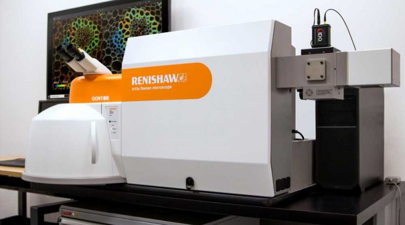
Renishaw is pleased to introduce fluorescence lifetime imaging microscopy (FLIM) functionality to its inVia™ confocal Raman microscope. In partnership with Becker & Hickl GmbH, the pioneers in time-correlated single photon counting (TCSPC), the system combines both Raman and FLIM in a single instrument. This integration can overlay both FLIM and Raman images with pixel-to-pixel correlation, thus increasing understanding of samples across a range of applications.
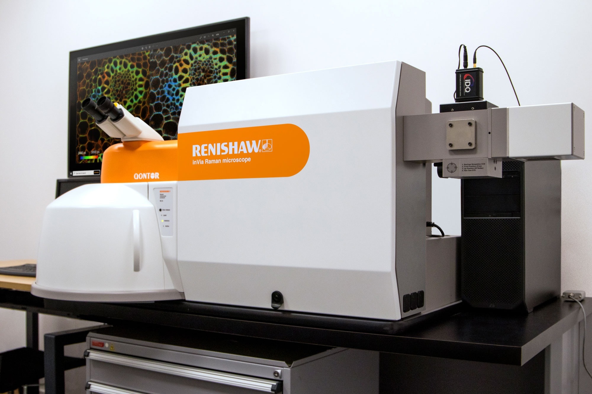
Image Credit: Renishaw
FLIM imaging
In the system, when light from a pulsed laser interacts with a fluorophore, the fluorescence lifetime is measured using an advanced detector and high-speed electronics. This fluorescence lifetime is the characteristic time a fluorophore remains in an excited electronic state before returning to the ground state by emitting a photon. The lifetime of fluorescence decay is sensitive to the molecular environment and the resulting changes in molecular conformation. The FLIM technique can be used to study a range of properties from pH/ion/oxygen concentration to molecular binding.
Combined FLIM with Raman microscopy
FLIM and Raman microscopy are complementary methods for label-free imaging of biological cells and tissue sections. FLIM provides insight into molecular interactions and can be performed at least 10× faster than Raman imaging. Raman spectroscopy boasts high chemical specificity and high-resolution chemical images. This combination of techniques is ideal for rapid identification of regions of interest by FLIM, followed by chemical and structural characterisation by Raman imaging.
For FLIM data analysis, levitra schmelztabletten beipackzettel comprehensive software from Becker & Hickl is included with multi-exponential, incomplete and shifted-component analysis models. The software also includes proprietary phasor analysis to distinguish lifetime populations. Fluorescence lifetime and Raman images can be directly overlaid due to the use of the highly accurate Renishaw MS30 high speed encoded stage for both imaging modalities.
“We are pleased to announce a further enhancement to our award-winning inVia Raman microscope. Our collaboration with the leaders in FLIM technology, Becker and Hickl, has produced a combined FLIM Raman microscope leading to exciting new application areas.”
David Reece, Renishaw Spectroscopy Business Development Manager
For further information on fluorescence lifetime imaging microscopy (FLIM): Fluorescence lifetime imaging microscopy (FLIM) (renishaw.com)
For further information on Renishaw Raman Spectroscopy: www.renishaw.com/raman
Source: Read Full Article