
nexium photo
Holotomography is a scientific method used to image cells in two- and three-dimensions without the need for sectioning or labeling with external proteins.
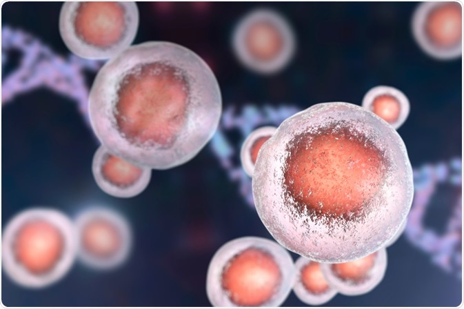 Image Credit: Kateryna Kon / Shutterstock
Principles of holotomography
Holotomography uses lasers to measure the refractive index in all three dimensions. As the refractive index of different objects inside a cell varies, this method can be used as a contrast agent to visualise different components inside a cell.
As light passes through an object, trazodone sleep time the phase of the light shifts based on its refractive index. Visualising both the incident and emergent light together can help in viewing the difference in the brightness, depending on how much the light has shifted. This property is used to view cells and tissues.
Three-dimensional refractive index
This method helps to infer the refractive index of an object in all dimensions. First, multiple two-dimensional tomographic images are taken at different angles. Subsequently, a 3D tomogram is reconstructed using these images.
Electron microscopy and laser scanning microscopes require extensive preparation of the sample and labelling. However, the basic principle of holotomography is similar to the phase contrast microscope where the contrast is generated due to the shift in the phase of light based on its refractive index. Thus, there is no need for fluorescent proteins, sectioning, or fixing the samples before imaging.
High resolution
The lateral resolution provided in this microscopy is around 166nm. This is the shortest distance between two objects inside a cell, which can be resolved using holotomography in the lateral direction. The resolution in the axial direction is 1 micron, and it provides a maximum field view of 80um.
Fast imaging two-and three-dimensional imaging
Holotomography has a fast scanning speed, and this property is useful when you are imaging live samples and live processes. The imaging speed can be up to 150 frames/sec for two-dimensional imaging and 2.5 frames/sec for three-dimensional imaging.
If the speed is slow, then it may not be able to catch cell processes that occur at a fast rate. Thus, it is critical to employ imaging methods which have a fast scanning speed while imaging live cellular processes.
Intact live cell imaging
This method can be used on cells without fixing, which kills the cells, or sectioning. Both of these processes require time and reagents. Also, killing the cell can also provide false results where the expression of genes and proteins may not be similar to living cells.
Some methods involve extracting the cellular contents from inside a cell and then visualising it. Such methods may also depict an altered picture of expression of genes, RNA, or proteins as the integrity of the cell is lost in this case. This method provides a way to observe inside a cell without killing or damaging its cellular integrity, thus, providing one of the best ways to visualise cells and their behaviour.
Quantitative imaging
Different methods have been used to quantify the properties of cells; however, these methods have limitations while assessing the cellular properties in three dimensions. Also, these methods require labeling proteins, such as green fluorescent proteins (GFP) or dyes. The inclusion of these factors can itself alter the quantitative parameters that are measured.
Quantitative methods that employ changes in phase properties inside a cell provides a non-invasive, unlabelled, and quick method to analyze cell behaviors. Using three-dimensional holotomography different quantitative parameters, such as refractive index, and local cytoplasmic concentration, as well as dry mass, distinguishing between cytoplasm and nuclei, can be derived.
Sources
- www.biorxiv.org/content/biorxiv/early/2017/10/20/206821.full.pdf
- http://www.tomocube.com/product/ht-1/
- https://www.nature.com/articles/s41598-018-24393-0
Further Reading
- All Holotomography Content
- What is Holotomography?
Last Updated: Oct 24, 2018

Written by
Dr. Surat P
Dr. Surat graduated with a Ph.D. in Cell Biology and Mechanobiology from the Tata Institute of Fundamental Research (Mumbai, India) in 2016. Prior to her Ph.D., Surat studied for a Bachelor of Science (B.Sc.) degree in Zoology, during which she was the recipient of anIndian Academy of SciencesSummer Fellowship to study the proteins involved in AIDs. She produces feature articles on a wide range of topics, such as medical ethics, data manipulation, pseudoscience and superstition, education, and human evolution. She is passionate about science communication and writes articles covering all areas of the life sciences.
Source: Read Full Article