
Insight into the Spinal Cord
The spinal cord is a long, cylindrical structure connecting the brain and body. It performs several vital neurophysiological functions, ranging from sensory signals processing to autonomic control of visceral functions.
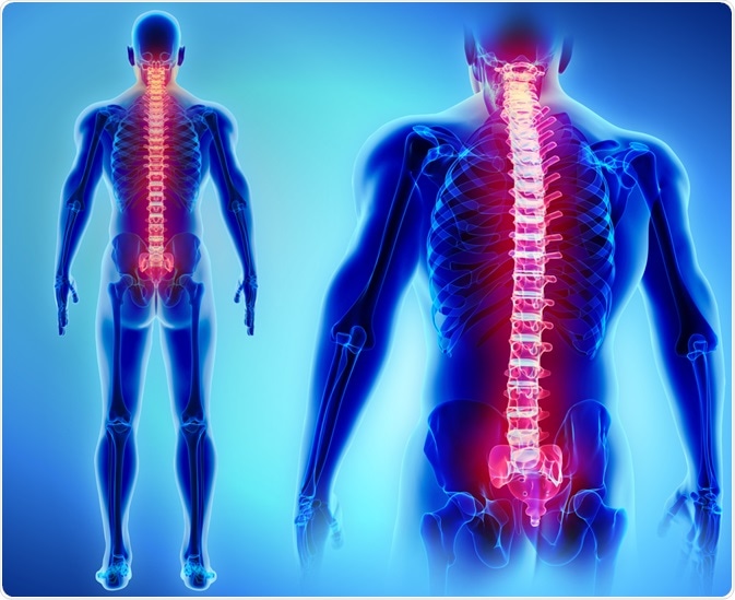
Image Credit: MDGRPHCS/Shutterstock.com
What is the anatomy of the spinal cord?
The human spinal cord is a cylindrical structure of nerve tissue that is protected by the spinal column and composed of uniformly organized white and grey matters. The spinal column is composed of bony structures called vertebrae.
The spinal cord is divided into 4 regions: cervical, thoracic, lumbar, and sacral regions. These regions are further divided into 31 segments: 8 cervical segments, 12 thoracic segments, 5 lumber segments, 5 sacral segments, and 1 coccygeal segment.
The length and diameter of the spinal cord are 40 – 50 cm and 1 – 1.5 cm, respectively. The structure is extended from the foramen magnum (situated at the base of the skull) to the lumbar vertebrae.
Two rows of nerve roots are present on each side of the spinal cord, which join distally to form 31 pairs of spinal nerves. Each pair of the spinal nerves leaves each spinal cord segment and innervates a dermatome, which is a particular skin region supplied by a single nerve fiber.
Each spinal nerve comprises motor and sensory nerve fibers. Receptors present in the skin transmit signals to the spinal cord via sensory nerve fibers, which enter the spinal cord via the dorsal root. Some of these nerve fibers form connections with other nerves in the dorsal horn, whereas some fibers directly continue up to the brain. The motor nerve fibers in the ventral horn send signals to the skeletal and smooth muscles via the ventral root to control voluntary and involuntary reflexes.
The spinal cord is surrounded by three meninges (membranes covering the brain and spinal cord): the dura mater, arachnoid mater, and pia mater. The dura mater forms the outermost covering, followed by the arachnoid mater and pia mater. The dura mater is separated from the spinal canal by the epidural space. The arachnoid mater is a delicate covering situated between the dura mater and the pia mater. It is separated from the pia mater by the subarachnoid space, which expands to form the lumbar cistern.
During a lumbar puncture, cerebrospinal fluid is obtained from this space. The pia mater is the thin innermost membrane that covers the spinal cord, nerve roots, and blood vessels. The pia mater expands between the nerve roots to form the denticulate ligaments, which connect to the dura mater to suspend the spinal cord in the spinal canal.
What are the spinal cord tracts?
There are two types of tracts: ascending and descending tracts.
The nerve fibers forming the ascending tract arise from the nerve cells present in the dorsal root ganglion. Sensory information related to temperature, pressure, touch, pain, vibration, etc., is transmitted from the skin receptors to the central nervous system via the ascending tracts. Unconscious proprioception information is also transmitted from the muscles and joints to the cerebellum via the ascending tracts.
Descending tracts, on the other hand, emerge from different cortical brain regions and brainstem nuclei. The descending tracts transmit information to control motor activities, including vascular and somatic reflexes, muscle tone, posture, and balance.
The spinal vasculature
Three longitudinal arteries supply the spinal cord: one anterior spinal artery and two posterior spinal arteries. Also, anterior and posterior segmental medullary arteries supply the spinal cord by entering through the nerve roots.
The venous blood circulation is carried out by three anterior and three posterior spinal veins. The spinal veins drain into the systemic segmental veins via the internal and external vertebral plexuses.
Any disruption in the spinal arterial blood supply can lead to nerve cell death, a condition medically termed as spinal infarction or spinal cord stroke. The condition is commonly caused by vertebral dislocation or fracture, compression due to tumors, blood vessel inflammation, or cholesterol accumulation in the arterial wall. Muscle weakness and paralysis are the common consequences of spinal cord infarction.
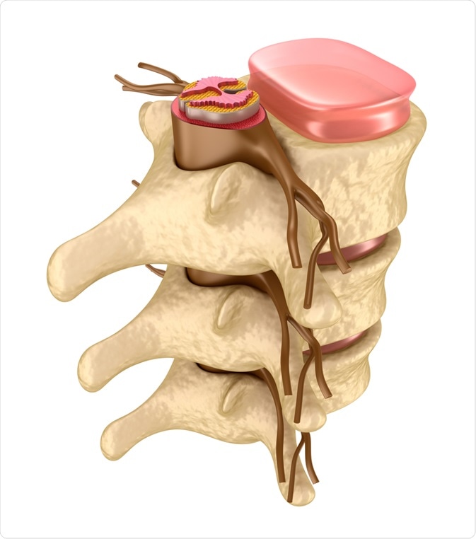
Image Credit: Alex Mit/Shutterstock.com
What are the functions of the spinal cord?
The spinal cord works in synergy with the brain to control important neurophysiological functions. While the brain works as a command center, the spinal cord works as a communicating pathway between the body and brain.
The peripheral nervous system is composed of nerve pairs that come out from the left and right sides of the spinal cord and spread throughout the body. A vast network created by these nerve strands establishes the connection between the body and brain to control various motor, sensory, and autonomic functions.
Voluntary movements of the muscles and joints are among the motor functions controlled by the spinal cord. The touch, pressure, pain, and temperature sensations are examples of sensory functions controlled by the spinal cord. Regarding autonomic functions, the spinal nerves regulate vital physiological functions, including heart rate, blood pressure, digestion, urination, and body temperature.
Sources
- The University of Texas. 2020. Anatomy of the spinal cord. https://nba.uth.tmc.edu/neuroscience/m/s2/chapter03.html
- TeachMeAnatomy. The spinal cord. https://teachmeanatomy.info/back/nerves/spinal-cord/
- The University of Alabama. 2020. What does the spinal cord do? www.uab.edu/…/what-does-the-spinal-cord-do
Last Updated: Oct 22, 2020

Written by
Dr. Sanchari Sinha Dutta
Dr. Sanchari Sinha Dutta is a science communicator who believes in spreading the power of science in every corner of the world. She has a Bachelor of Science (B.Sc.) degree and a Master's of Science (M.Sc.) in biology and human physiology. Following her Master's degree, Sanchari went on to study a Ph.D. in human physiology. She has authored more than 10 original research articles, all of which have been published in world renowned international journals.
Source: Read Full Article